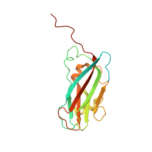
8BI4

Helical shell of CCMV capsid protein on DNA origami 6HB-2k


Experimental Data Snapshot

  • Method: ELECTRON MICROSCOPY
  • Resolution: 4.30 Å
  • Aggregation State: FILAMENT 
  • Reconstruction Method: SINGLE PARTICLE 

wwPDB Validation   3D Report Full Report


This is version 1.2 of the entry. See complete history


Literature

DNA-origami-directed virus capsid polymorphism.

Seitz, I.Saarinen, S.Kumpula, E.P.McNeale, D.Anaya-Plaza, E.Lampinen, V.Hytonen, V.P.Sainsbury, F.Cornelissen, J.J.L.M.Linko, V.Huiskonen, J.T.Kostiainen, M.A.

(2023) Nat Nanotechnol 18: 1205-1212

  • DOI: https://doi.org/10.1038/s41565-023-01443-x
  • Primary Citation of Related Structures:  
    8BI4

  • PubMed Abstract: 

    Viral capsids can adopt various geometries, most iconically characterized by icosahedral or helical symmetries. Importantly, precise control over the size and shape of virus capsids would have advantages in the development of new vaccines and delivery systems. However, current tools to direct the assembly process in a programmable manner are exceedingly elusive. Here we introduce a modular approach by demonstrating DNA-origami-directed polymorphism of single-protein subunit capsids. We achieve control over the capsid shape, size and topology by employing user-defined DNA origami nanostructures as binding and assembly platforms, which are efficiently encapsulated within the capsid. Furthermore, the obtained viral capsid coatings can shield the encapsulated DNA origami from degradation. Our approach is, moreover, not limited to a single type of capsomers and can also be applied to RNA-DNA origami structures to pave way for next-generation cargo protection and targeting strategies.


  • Organizational Affiliation

    Department of Bioproducts and Biosystems, Aalto University, Aalto, Finland.


Macromolecules
Find similar proteins by:  (by identity cutoff)  |  3D Structure
Entity ID: 1
MoleculeChains Sequence LengthOrganismDetailsImage
Coat protein
A, B, C, D, E
A, B, C, D, E, F
190Cowpea chlorotic mottle virusMutation(s): 0 
UniProt
Find proteins for P03601 (Cowpea chlorotic mottle virus)
Explore P03601 
Go to UniProtKB:  P03601
Entity Groups  
Sequence Clusters30% Identity50% Identity70% Identity90% Identity95% Identity100% Identity
UniProt GroupP03601
Sequence Annotations
Expand
  • Reference Sequence
Experimental Data & Validation

Experimental Data

  • Method: ELECTRON MICROSCOPY
  • Resolution: 4.30 Å
  • Aggregation State: FILAMENT 
  • Reconstruction Method: SINGLE PARTICLE 
EM Software:
TaskSoftware PackageVersion
RECONSTRUCTIONcryoSPARC3.3.2
MODEL REFINEMENTPHENIX1.19

Structure Validation

View Full Validation Report


Entry History & Funding Information

Deposition Data


Funding OrganizationLocationGrant Number
European Research Council (ERC)European Union101002258
Academy of FinlandFinland341057

Revision History  (Full details and data files)

  • Version 1.0: 2023-07-05
    Type: Initial release
  • Version 1.1: 2023-07-26
    Changes: Database references
  • Version 1.2: 2023-10-25
    Changes: Data collection, Database references