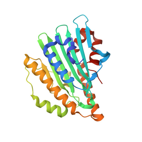
7YK9

Neutron Structure of PcyA I86D Mutant Complexed with Biliverdin at Room Temperature


Experimental Data Snapshot

  • Method: X-RAY DIFFRACTION
  • Resolution: 1.90 Å
  • R-Value Free: 0.174 
  • R-Value Work: 0.138 
  • R-Value Observed: 0.141 

  • Method: NEUTRON DIFFRACTION
  • Resolution: 2.00 Å
  • R-Value Free: 0.203 
  • R-Value Work: 0.171 

Starting Model: experimental
View more details

wwPDB Validation   3D Report Full Report


This is version 1.1 of the entry. See complete history


Literature

Neutron crystallography and quantum chemical analysis of bilin reductase PcyA mutants reveal substrate and catalytic residue protonation states.

Joutsuka, T.Nanasawa, R.Igarashi, K.Horie, K.Sugishima, M.Hagiwara, Y.Wada, K.Fukuyama, K.Yano, N.Mori, S.Ostermann, A.Kusaka, K.Unno, M.

(2022) J Biol Chem 299: 102763-102763

  • DOI: https://doi.org/10.1016/j.jbc.2022.102763
  • Primary Citation of Related Structures:  
    7YK9, 7YKB

  • PubMed Abstract: 

    PcyA, a ferredoxin-dependent bilin pigment reductase, catalyzes the site-specific reduction of the two vinyl groups of biliverdin (BV), producing phycocyanobilin. Previous neutron crystallography detected both the neutral BV and its protonated form (BVH + ) in the wildtype (WT) PcyA-BV complex, and a nearby catalytic residue Asp105 was found to have two conformations (protonated and deprotonated). Semiempirical calculations have suggested that the protonation states of BV are reflected in the absorption spectrum of the WT PcyA-BV complex. In the previously determined absorption spectra of the PcyA D105N and I86D mutants, complexed with BV, a peak at 730 nm, observed in the WT, disappeared and increased, respectively. Here, we performed neutron crystallography and quantum chemical analysis of the D105N-BV and I86D-BV complexes to determine the protonation states of BV and the surrounding residues and study the correlation between the absorption spectra and protonation states around BV. Neutron structures elucidated that BV in the D105N mutant is in a neutral state, whereas that in the I86D mutant is dominantly in a protonated state. Glu76 and His88 showed different hydrogen bonding with surrounding residues compared with WT PcyA, further explaining why D105N and I86D have much lower activities for phycocyanobilin synthesis than the WT PcyA. Our quantum mechanics/molecular mechanics calculations of the absorption spectra showed that the spectral change in D105N arises from Glu76 deprotonation, consistent with the neutron structure. Collectively, our findings reveal more mechanistic details of bilin pigment biosynthesis.


  • Organizational Affiliation

    Graduate School of Science and Engineering, Ibaraki University, Hitachi, Ibaraki, Japan; Frontier Research Center for Applied Atomic Sciences, Ibaraki University, Naka-Tokai, Ibaraki, Japan. Electronic address: tatsuya.joutsuka.joe@vc.ibaraki.ac.jp.


Macromolecules
Find similar proteins by:  (by identity cutoff)  |  3D Structure
Entity ID: 1
MoleculeChains Sequence LengthOrganismDetailsImage
Phycocyanobilin:ferredoxin oxidoreductase248Synechocystis sp. PCC 6803Mutation(s): 1 
Gene Names: pcyA
EC: 1.3.7.5
UniProt
Find proteins for Q55891 (Synechocystis sp. (strain ATCC 27184 / PCC 6803 / Kazusa))
Explore Q55891 
Go to UniProtKB:  Q55891
Entity Groups  
Sequence Clusters30% Identity50% Identity70% Identity90% Identity95% Identity100% Identity
UniProt GroupQ55891
Sequence Annotations
Expand
  • Reference Sequence
Small Molecules
Ligands 1 Unique
IDChains Name / Formula / InChI Key2D Diagram3D Interactions
BLR (Subject of Investigation/LOI)
Query on BLR

Download Ideal Coordinates CCD File 
B [auth A]3-[5-[(Z)-(4-ethenyl-3-methyl-5-oxidanylidene-pyrrol-2-ylidene)methyl]-2-[[5-[(Z)-(3-ethenyl-4-methyl-5-oxidanylidene-pyrrol-2-ylidene)methyl]-3-(3-hydroxy-3-oxopropyl)-4-methyl-1H-pyrrol-2-yl]methyl]-4-methyl-1H-pyrrol-3-yl]propanoic acid
C33 H36 N4 O6
BPYKTIZUTYGOLE-IFADSCNNSA-N
Experimental Data & Validation

Experimental Data

  • Method: X-RAY DIFFRACTION
  • Resolution: 1.90 Å
  • R-Value Free: 0.174 
  • R-Value Work: 0.138 
  • R-Value Observed: 0.141 
  • Space Group: P 21 21 2
  • Method: NEUTRON DIFFRACTION
  • Resolution: 2.00 Å
  • R-Value Free: 0.203 
  • R-Value Work: 0.171 
  • Space Group: P 21 21 2
Unit Cell:
Length ( Å )Angle ( ˚ )
a = 71.325α = 90
b = 97.415β = 90
c = 43.134γ = 90
Software Package:
Software NamePurpose
PHENIXrefinement
HKL-2000data reduction
HKL-2000data scaling
REFMACphasing

Structure Validation

View Full Validation Report


Entry History & Funding Information

Deposition Data


Funding OrganizationLocationGrant Number
Japan Society for the Promotion of Science (JSPS)Japan16K07261

Revision History  (Full details and data files)

  • Version 1.0: 2023-01-25
    Type: Initial release
  • Version 1.1: 2024-04-03
    Changes: Data collection, Refinement description