7X32

Crystal structure of E. coli NfsB in complex with berberine


Experimental Data Snapshot

  • Method: X-RAY DIFFRACTION
  • Resolution: 1.83 Å
  • R-Value Free: 0.241 
  • R-Value Work: 0.205 
  • R-Value Observed: 0.206 

Starting Model: experimental
View more details

wwPDB Validation   3D Report Full Report


Ligand Structure Quality Assessment 


This is version 1.1 of the entry. See complete history


Literature

Structural basis for the transformation of the traditional medicine berberine by bacterial nitroreductase.

Wen, H.Y.Pan, L.B.Ma, S.R.Yang, X.Y.Hu, J.C.Zhao, H.F.Gao, Z.Q.Dong, Y.H.Wang, Y.Zhang, H.

(2022) Acta Crystallogr D Struct Biol 78: 1273-1282

  • DOI: https://doi.org/10.1107/S2059798322008373
  • Primary Citation of Related Structures:  
    7X32

  • PubMed Abstract: 

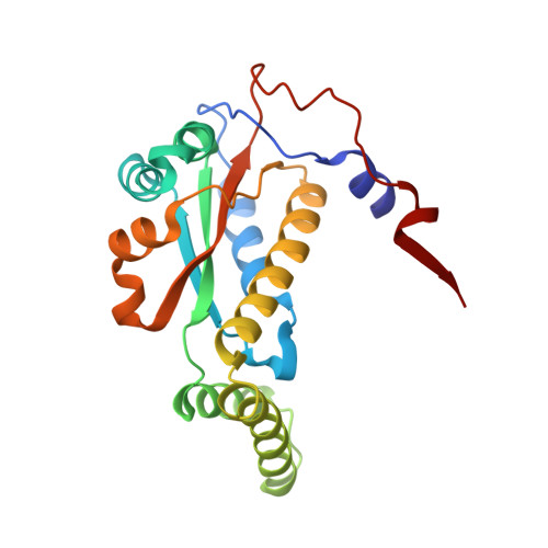
    The bacterial nitroreductases (NRs) NfsB and NfsA are conserved homodimeric FMN-dependent flavoproteins that are responsible for the reduction of nitroaromatic substrates. Berberine (BBR) is a plant-derived isoquinoline alkaloid with a large conjugated ring system that is widely used in the treatment of various diseases. It was recently found that the gut microbiota convert BBR into dihydroberberine (dhBBR, the absorbable form) mediated by bacterial NRs. The molecular basis for the transformation of BBR by the gut microbiota remains unclear. Here, kinetic studies showed that NfsB from Escherichia coli (EcNfsB), rather than EcNfsA, is responsible for the conversion of BBR to dhBBR in spite of a low reaction rate. The crystal structure of the EcNfsB-BBR complex showed that BBR binds into the active pocket at the dimer interface, and its large conjugated plane stacks above the plane of the FMN cofactor in a nearly parallel orientation. BBR is mainly stabilized by π-stacking interactions with both neighboring aromatic residues and FMN. Structure-based mutagenesis studies further revealed that the highly conserved Phe70 and Phe199 are important residues for the conversion of BBR. The structure revealed that the C6 atom of BBR (which receives the hydride) is ∼7.5 Å from the N5 atom of FMN (which donates the hydride), which is too distant for hydride transfer. Notably, several well ordered water molecules make hydrogen-bond/van der Waals contacts with the N1 atom of BBR in the active site, which probably donate protons in conjunction with electron transfer from FMN. The structure-function studies revealed the mechanism for the recognition and binding of BBR by bacterial NRs and may help to understand the conversion of BBR by the gut microbiota.


  • Organizational Affiliation

    School of Life Sciences, University of Science and Technology of China, Hefei 230027, People's Republic of China.


Macromolecules
Find similar proteins by:  (by identity cutoff)  |  3D Structure
Entity ID: 1
MoleculeChains Sequence LengthOrganismDetailsImage
Dihydropteridine reductase
A, B, C, D, E
A, B, C, D, E, F, G, H
217Escherichia coliMutation(s): 0 
Gene Names: 
EC: 1.5.1.34 (PDB Primary Data), 1 (PDB Primary Data)
UniProt
Find proteins for P38489 (Escherichia coli (strain K12))
Explore P38489 
Go to UniProtKB:  P38489
Entity Groups  
Sequence Clusters30% Identity50% Identity70% Identity90% Identity95% Identity100% Identity
UniProt GroupP38489
Sequence Annotations
Expand
  • Reference Sequence
Small Molecules
Ligands 4 Unique
IDChains Name / Formula / InChI Key2D Diagram3D Interactions
FMN (Subject of Investigation/LOI)
Query on FMN

Download Ideal Coordinates CCD File 
AA [auth H]
I [auth A]
K [auth B]
M [auth C]
P [auth D]
AA [auth H],
I [auth A],
K [auth B],
M [auth C],
P [auth D],
T [auth E],
W [auth F],
X [auth G]
FLAVIN MONONUCLEOTIDE
C17 H21 N4 O9 P
FVTCRASFADXXNN-SCRDCRAPSA-N
BER (Subject of Investigation/LOI)
Query on BER

Download Ideal Coordinates CCD File 
BA [auth H],
N [auth C],
Q [auth D],
U [auth E],
Y [auth G]
BERBERINE
C20 H18 N O4
YBHILYKTIRIUTE-UHFFFAOYSA-N
GOL (Subject of Investigation/LOI)
Query on GOL

Download Ideal Coordinates CCD File 
CA [auth H],
J [auth A],
R [auth D]
GLYCEROL
C3 H8 O3
PEDCQBHIVMGVHV-UHFFFAOYSA-N
DMS (Subject of Investigation/LOI)
Query on DMS

Download Ideal Coordinates CCD File 
DA [auth H]
L [auth B]
O [auth C]
S [auth D]
V [auth E]
DA [auth H],
L [auth B],
O [auth C],
S [auth D],
V [auth E],
Z [auth G]
DIMETHYL SULFOXIDE
C2 H6 O S
IAZDPXIOMUYVGZ-UHFFFAOYSA-N
Experimental Data & Validation

Experimental Data

  • Method: X-RAY DIFFRACTION
  • Resolution: 1.83 Å
  • R-Value Free: 0.241 
  • R-Value Work: 0.205 
  • R-Value Observed: 0.206 
  • Space Group: P 1
Unit Cell:
Length ( Å )Angle ( ˚ )
a = 57.466α = 74.05
b = 77.941β = 79.44
c = 116.128γ = 74.26
Software Package:
Software NamePurpose
PHENIXrefinement
PDB_EXTRACTdata extraction
XDSdata reduction
XDSdata scaling
PHASERphasing

Structure Validation

View Full Validation Report


Ligand Structure Quality Assessment 


Entry History & Funding Information

Deposition Data


Funding OrganizationLocationGrant Number
National Natural Science Foundation of China (NSFC)China31970152

Revision History  (Full details and data files)

  • Version 1.0: 2022-10-12
    Type: Initial release
  • Version 1.1: 2023-11-29
    Changes: Data collection, Refinement description