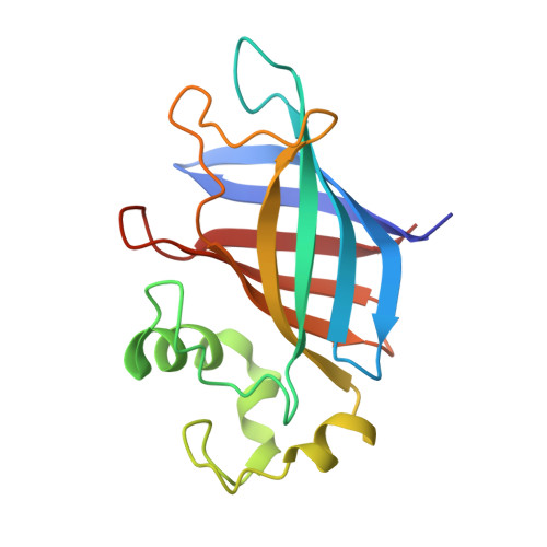
7U3F

GID4 in complex with compound 4


Experimental Data Snapshot

  • Method: X-RAY DIFFRACTION
  • Resolution: 2.30 Å
  • R-Value Free: 0.262 
  • R-Value Work: 0.233 
  • R-Value Observed: 0.235 

Starting Model: experimental
View more details

wwPDB Validation   3D Report Full Report


Ligand Structure Quality Assessment 


This is version 1.2 of the entry. See complete history


Literature

Discovery and Structural Characterization of Small Molecule Binders of the Human CTLH E3 Ligase Subunit GID4.

Chana, C.K.Maisonneuve, P.Posternak, G.Grinberg, N.G.A.Poirson, J.Ona, S.M.Ceccarelli, D.F.Mader, P.St-Cyr, D.J.Pau, V.Kurinov, I.Tang, X.Deng, D.Cui, W.Su, W.Kuai, L.Soll, R.Tyers, M.Rost, H.L.Batey, R.A.Taipale, M.Gingras, A.C.Sicheri, F.

(2022) J Med Chem 65: 12725-12746

  • DOI: https://doi.org/10.1021/acs.jmedchem.2c00509
  • Primary Citation of Related Structures:  
    7U3E, 7U3F, 7U3G, 7U3H, 7U3I, 7U3J, 7U3K, 7U3L

  • PubMed Abstract: 

    Targeted protein degradation (TPD) strategies exploit bivalent small molecules to bridge substrate proteins to an E3 ubiquitin ligase to induce substrate degradation. Few E3s have been explored as degradation effectors due to a dearth of E3-binding small molecules. We show that genetically induced recruitment to the GID4 subunit of the CTLH E3 complex induces protein degradation. An NMR-based fragment screen followed by structure-guided analog elaboration identified two binders of GID4, 16 and 67 , with K d values of 110 and 17 μM in vitro . A parallel DNA-encoded library (DEL) screen identified five binders of GID4, the best of which, 88 , had a K d of 5.6 μM in vitro and an EC 50 of 558 nM in cells with strong selectivity for GID4. X-ray co-structure determination revealed the basis for GID4-small molecule interactions. These results position GID4-CTLH as an E3 for TPD and provide candidate scaffolds for high-affinity moieties that bind GID4.


  • Organizational Affiliation

    Lunenfeld-Tanenbaum Research Institute, Sinai Health, Toronto, Ontario M5G 1X5, Canada.


Macromolecules
Find similar proteins by:  (by identity cutoff)  |  3D Structure
Entity ID: 1
MoleculeChains Sequence LengthOrganismDetailsImage
Glucose-induced degradation protein 4 homolog167Homo sapiensMutation(s): 0 
Gene Names: GID4C17orf39VID24
UniProt & NIH Common Fund Data Resources
Find proteins for Q8IVV7 (Homo sapiens)
Explore Q8IVV7 
Go to UniProtKB:  Q8IVV7
PHAROS:  Q8IVV7
GTEx:  ENSG00000141034 
Entity Groups  
Sequence Clusters30% Identity50% Identity70% Identity90% Identity95% Identity100% Identity
UniProt GroupQ8IVV7
Sequence Annotations
Expand
  • Reference Sequence
Small Molecules
Experimental Data & Validation

Experimental Data

  • Method: X-RAY DIFFRACTION
  • Resolution: 2.30 Å
  • R-Value Free: 0.262 
  • R-Value Work: 0.233 
  • R-Value Observed: 0.235 
  • Space Group: P 21 21 21
Unit Cell:
Length ( Å )Angle ( ˚ )
a = 40.34α = 90
b = 57.28β = 90
c = 72.53γ = 90
Software Package:
Software NamePurpose
PHENIXrefinement
XDSdata reduction
XSCALEdata scaling
PHASERphasing
PDB_EXTRACTdata extraction

Structure Validation

View Full Validation Report


Ligand Structure Quality Assessment 


Entry History & Funding Information

Deposition Data


Funding OrganizationLocationGrant Number
Canadian Institutes of Health Research (CIHR)CanadaFDN-143277

Revision History  (Full details and data files)

  • Version 1.0: 2022-10-05
    Type: Initial release
  • Version 1.1: 2022-10-26
    Changes: Database references
  • Version 1.2: 2023-10-18
    Changes: Data collection, Refinement description