7STZ

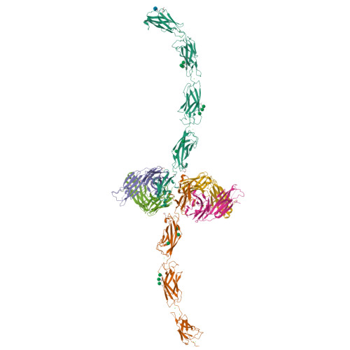
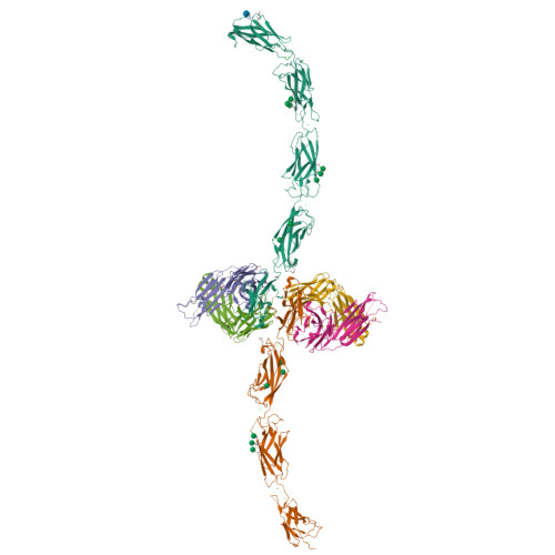
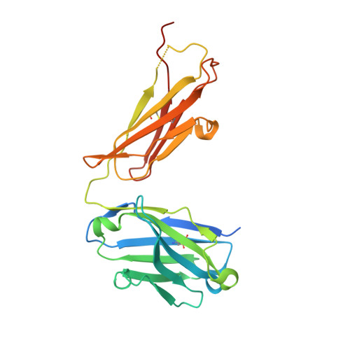
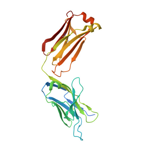
Crystal Structure of Human E-cadherin EC1-5 bound by mouse monoclonal antibody Fab mAb-1_19A11


Experimental Data Snapshot

  • Method: X-RAY DIFFRACTION
  • Resolution: 2.95 Å
  • R-Value Free: 0.208 
  • R-Value Work: 0.182 
  • R-Value Observed: 0.182 

Starting Model: experimental
View more details

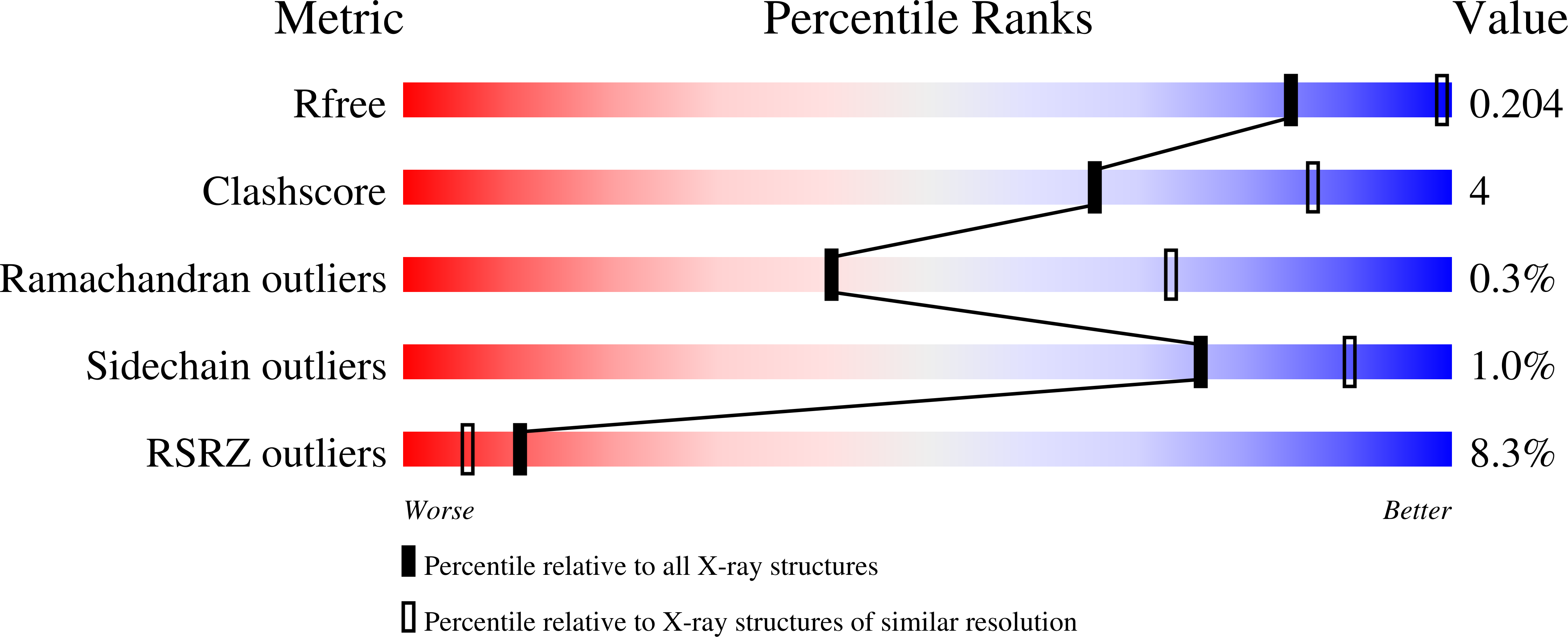
wwPDB Validation   3D Report Full Report


Ligand Structure Quality Assessment 


This is version 1.3 of the entry. See complete history


Literature

Regulation of multiple dimeric states of E-cadherin by adhesion activating antibodies revealed through Cryo-EM and X-ray crystallography.

Maker, A.Bolejack, M.Schecterson, L.Hammerson, B.Abendroth, J.Edwards, T.E.Staker, B.Myler, P.J.Gumbiner, B.M.

(2022) PNAS Nexus 1: pgac163-pgac163

  • DOI: https://doi.org/10.1093/pnasnexus/pgac163
  • Primary Citation of Related Structures:  
    6VEL, 7STZ

  • PubMed Abstract: 

    E-cadherin adhesion is regulated at the cell surface, a process that can be replicated by activating antibodies. We use cryo-electron microscopy (EM) and X-ray crystallography to examine functional states of the cadherin adhesive dimer. This dimer is mediated by N-terminal beta strand-swapping involving Trp2, and forms via a different transient X-dimer intermediate. X-dimers are observed in cryo-EM along with monomers and strand-swap dimers, indicating that X-dimers form stable interactions. A novel EC4-mediated dimer was also observed. Activating Fab binding caused no gross structural changes in E-cadherin monomers, but can facilitate strand swapping. Moreover, activating Fab binding is incompatible with the formation of the X-dimer. Both cryo-EM and X-ray crystallography reveal a distinctive twisted strand-swap dimer conformation caused by an outward shift in the N-terminal beta strand that may represent a strengthened state. Thus, regulation of adhesion involves changes in cadherin dimer configurations.


  • Organizational Affiliation

    Department of Biochemistry, University of Washington, USA.


Macromolecules
Find similar proteins by:  (by identity cutoff)  |  3D Structure
Entity ID: 1
MoleculeChains Sequence LengthOrganismDetailsImage
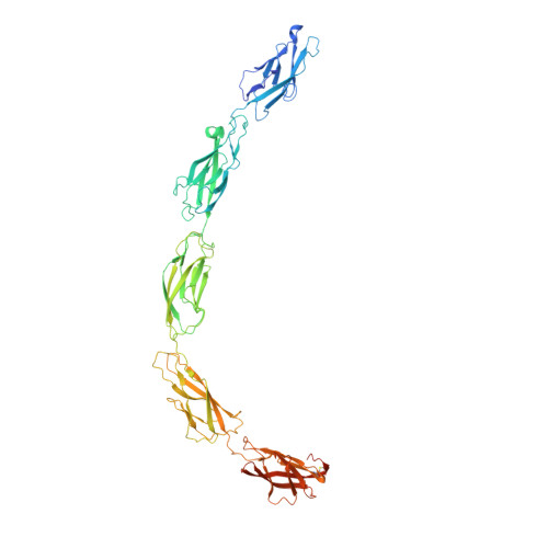
Cadherin-1A [auth C],
D
590Homo sapiensMutation(s): 0 
Gene Names: CDH1CDHEUVO
UniProt & NIH Common Fund Data Resources
Find proteins for P12830 (Homo sapiens)
Explore P12830 
Go to UniProtKB:  P12830
PHAROS:  P12830
GTEx:  ENSG00000039068 
Entity Groups  
Sequence Clusters30% Identity50% Identity70% Identity90% Identity95% Identity100% Identity
UniProt GroupP12830
Glycosylation
Glycosylation Sites: 4Go to GlyGen: P12830-1
Sequence Annotations
Expand
  • Reference Sequence
Find similar proteins by:  (by identity cutoff)  |  3D Structure
Entity ID: 2
MoleculeChains Sequence LengthOrganismDetailsImage
mAb-1_19A11 Heavy ChainB [auth H],
E [auth I]
246Mus musculusMutation(s): 0 
Gene Names: Igh
Entity Groups  
Sequence Clusters30% Identity50% Identity70% Identity90% Identity95% Identity100% Identity
Sequence Annotations
Expand
  • Reference Sequence
Find similar proteins by:  (by identity cutoff)  |  3D Structure
Entity ID: 3
MoleculeChains Sequence LengthOrganismDetailsImage
mAb-1_19A11 Light ChainC [auth L],
F [auth M]
240Mus musculusMutation(s): 0 
Gene Names: LC
Entity Groups  
Sequence Clusters30% Identity50% Identity70% Identity90% Identity95% Identity100% Identity
Sequence Annotations
Expand
  • Reference Sequence
Small Molecules
Ligands 6 Unique
IDChains Name / Formula / InChI Key2D Diagram3D Interactions
NAG
Query on NAG

Download Ideal Coordinates CCD File 
H [auth C]2-acetamido-2-deoxy-beta-D-glucopyranose
C8 H15 N O6
OVRNDRQMDRJTHS-FMDGEEDCSA-N
PG4
Query on PG4

Download Ideal Coordinates CCD File 
CB [auth I],
HA [auth H],
I [auth C],
IA [auth D],
J [auth C]
TETRAETHYLENE GLYCOL
C8 H18 O5
UWHCKJMYHZGTIT-UHFFFAOYSA-N
BMA
Query on BMA

Download Ideal Coordinates CCD File 
JA [auth D]
KA [auth D]
L [auth C]
LA [auth D]
MA [auth D]
JA [auth D],
KA [auth D],
L [auth C],
LA [auth D],
MA [auth D],
N [auth C],
NA [auth D],
O [auth C],
OA [auth D],
P [auth C],
R [auth C],
S [auth C]
beta-D-mannopyranose
C6 H12 O6
WQZGKKKJIJFFOK-RWOPYEJCSA-N
MAN
Query on MAN

Download Ideal Coordinates CCD File 
K [auth C],
M [auth C],
Q [auth C]
alpha-D-mannopyranose
C6 H12 O6
WQZGKKKJIJFFOK-PQMKYFCFSA-N
EDO
Query on EDO

Download Ideal Coordinates CCD File 
AB [auth I],
BB [auth I],
FA [auth H],
G [auth C],
GA [auth H]
1,2-ETHANEDIOL
C2 H6 O2
LYCAIKOWRPUZTN-UHFFFAOYSA-N
CA
Query on CA

Download Ideal Coordinates CCD File 
AA [auth C]
BA [auth C]
CA [auth C]
DA [auth C]
DB [auth M]
AA [auth C],
BA [auth C],
CA [auth C],
DA [auth C],
DB [auth M],
EA [auth C],
PA [auth D],
QA [auth D],
RA [auth D],
SA [auth D],
T [auth C],
TA [auth D],
U [auth C],
UA [auth D],
V [auth C],
VA [auth D],
W [auth C],
WA [auth D],
X [auth C],
XA [auth D],
Y [auth C],
YA [auth D],
Z [auth C],
ZA [auth D]
CALCIUM ION
Ca
BHPQYMZQTOCNFJ-UHFFFAOYSA-N
Experimental Data & Validation

Experimental Data

  • Method: X-RAY DIFFRACTION
  • Resolution: 2.95 Å
  • R-Value Free: 0.208 
  • R-Value Work: 0.182 
  • R-Value Observed: 0.182 
  • Space Group: P 1 21 1
Unit Cell:
Length ( Å )Angle ( ˚ )
a = 85.34α = 90
b = 131.62β = 100.86
c = 201.76γ = 90
Software Package:
Software NamePurpose
XDSdata reduction
XSCALEdata scaling
PHENIXrefinement
PDB_EXTRACTdata extraction
PHASERphasing

Structure Validation

View Full Validation Report


Ligand Structure Quality Assessment 


Entry History & Funding Information

Deposition Data


Funding OrganizationLocationGrant Number
National Institutes of Health/National Institute Of Allergy and Infectious Diseases (NIH/NIAID)United States--

Revision History  (Full details and data files)

  • Version 1.0: 2022-01-26
    Type: Initial release
  • Version 1.1: 2023-02-01
    Changes: Database references
  • Version 1.2: 2023-10-25
    Changes: Data collection, Refinement description
  • Version 1.3: 2024-11-06
    Changes: Structure summary