7SR9

Human alpha-thrombin with 180- and 220- loops replaced with homologous loops from protein C


Experimental Data Snapshot

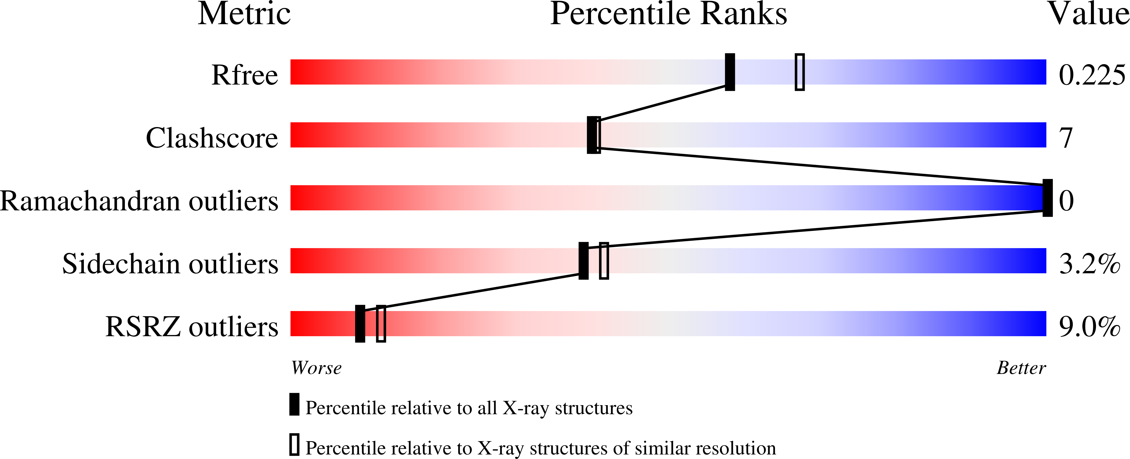
  • Method: X-RAY DIFFRACTION
  • Resolution: 2.10 Å
  • R-Value Free: 0.227 
  • R-Value Work: 0.178 
  • R-Value Observed: 0.181 

Starting Model: experimental
View more details

wwPDB Validation   3D Report Full Report


Ligand Structure Quality Assessment 


This is version 1.3 of the entry. See complete history


Literature

The active site region plays a critical role in Na + binding to thrombin.

Pelc, L.A.Koester, S.K.Kukla, C.R.Chen, Z.Di Cera, E.

(2022) J Biol Chem 298: 101458-101458

  • DOI: https://doi.org/10.1016/j.jbc.2021.101458
  • Primary Citation of Related Structures:  
    7SR9

  • PubMed Abstract: 

    The catalytic activity of thrombin and other enzymes of the blood coagulation and complement cascades is enhanced significantly by binding of Na + to a site >15 Å away from the catalytic residue S195, buried within the 180 and 220 loops that also contribute to the primary specificity of the enzyme. Rapid kinetics support a binding mechanism of conformational selection where the Na + -binding site is in equilibrium between open (N) and closed (N ) forms and the cation binds selectively to the N form. Allosteric transduction of this binding step produces enhanced catalytic activity. Molecular details on how Na + gains access to this site and communicates allosterically with the active site remain poorly defined. In this study, we show that the rate of the N →N transition is strongly correlated with the analogous E →E transition that governs the interaction of synthetic and physiologic substrates with the active site. This correlation supports the active site as the likely point of entry for Na + to its binding site. Mutagenesis and structural data rule out an alternative path through the pore defined by the 180 and 220 loops. We suggest that the active site communicates allosterically with the Na + site through a network of H-bonded water molecules that embeds the primary specificity pocket. Perturbation of the mobility of S195 and its H-bonding capabilities alters interaction with this network and influences the kinetics of Na + binding and allosteric transduction. These findings have general mechanistic relevance for Na + -activated proteases and allosteric enzymes.


  • Organizational Affiliation

    Edward A. Doisy Department of Biochemistry and Molecular Biology, Saint Louis University School of Medicine, St. Louis, Missouri, USA.


Macromolecules
Find similar proteins by:  (by identity cutoff)  |  3D Structure
Entity ID: 1
MoleculeChains Sequence LengthOrganismDetailsImage
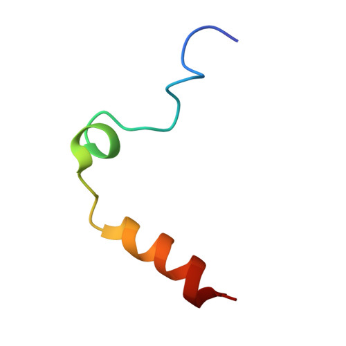
Thrombin light chain35Homo sapiensMutation(s): 0 
Gene Names: F2
EC: 3.4.21.5
UniProt & NIH Common Fund Data Resources
Find proteins for P00734 (Homo sapiens)
Explore P00734 
Go to UniProtKB:  P00734
PHAROS:  P00734
GTEx:  ENSG00000180210 
Entity Groups  
Sequence Clusters30% Identity50% Identity70% Identity90% Identity95% Identity100% Identity
UniProt GroupP00734
Sequence Annotations
Expand
  • Reference Sequence
Find similar proteins by:  (by identity cutoff)  |  3D Structure
Entity ID: 2
MoleculeChains Sequence LengthOrganismDetailsImage
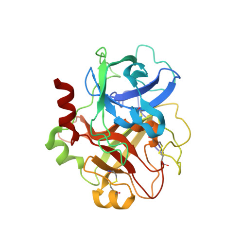
Thrombin heavy chain253Homo sapiensMutation(s): 9 
Gene Names: F2
EC: 3.4.21.5
UniProt & NIH Common Fund Data Resources
Find proteins for P00734 (Homo sapiens)
Explore P00734 
Go to UniProtKB:  P00734
PHAROS:  P00734
GTEx:  ENSG00000180210 
Entity Groups  
Sequence Clusters30% Identity50% Identity70% Identity90% Identity95% Identity100% Identity
UniProt GroupP00734
Glycosylation
Glycosylation Sites: 1Go to GlyGen: P00734-1
Sequence Annotations
Expand
  • Reference Sequence
Small Molecules
Ligands 3 Unique
IDChains Name / Formula / InChI Key2D Diagram3D Interactions
NAG
Query on NAG

Download Ideal Coordinates CCD File 
D [auth B]2-acetamido-2-deoxy-beta-D-glucopyranose
C8 H15 N O6
OVRNDRQMDRJTHS-FMDGEEDCSA-N
SO4
Query on SO4

Download Ideal Coordinates CCD File 
C [auth A],
E [auth B],
F [auth B],
G [auth B],
H [auth B]
SULFATE ION
O4 S
QAOWNCQODCNURD-UHFFFAOYSA-L
GOL
Query on GOL

Download Ideal Coordinates CCD File 
I [auth B]GLYCEROL
C3 H8 O3
PEDCQBHIVMGVHV-UHFFFAOYSA-N
Experimental Data & Validation

Experimental Data

  • Method: X-RAY DIFFRACTION
  • Resolution: 2.10 Å
  • R-Value Free: 0.227 
  • R-Value Work: 0.178 
  • R-Value Observed: 0.181 
  • Space Group: C 1 2 1
Unit Cell:
Length ( Å )Angle ( ˚ )
a = 98.969α = 90
b = 78.525β = 103.22
c = 49.602γ = 90
Software Package:
Software NamePurpose
REFMACrefinement
HKL-2000data reduction
HKL-2000data scaling
HKL2Mapphasing

Structure Validation

View Full Validation Report


Ligand Structure Quality Assessment 


Entry History & Funding Information

Deposition Data


Funding OrganizationLocationGrant Number
National Institutes of Health/National Heart, Lung, and Blood Institute (NIH/NHLBI)United States--

Revision History  (Full details and data files)

  • Version 1.0: 2021-12-08
    Type: Initial release
  • Version 1.1: 2022-06-22
    Changes: Database references
  • Version 1.2: 2023-10-18
    Changes: Data collection, Refinement description
  • Version 1.3: 2024-11-06
    Changes: Structure summary