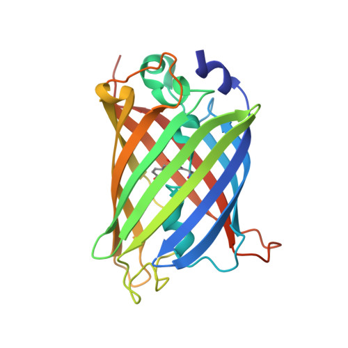
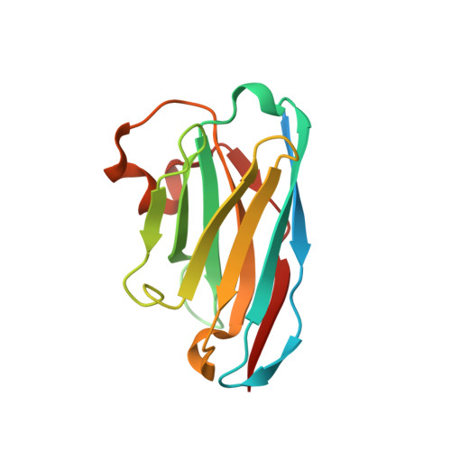
7SAH

Crystal Structure of LaG16 Nanobody bound to eGFP


Experimental Data Snapshot

  • Method: X-RAY DIFFRACTION
  • Resolution: 1.60 Å
  • R-Value Free: 0.177 
  • R-Value Work: 0.156 
  • R-Value Observed: 0.157 

Starting Model: experimental
View more details

wwPDB Validation   3D Report Full Report


This is version 2.1 of the entry. See complete history


Literature

High-efficiency recombinant protein purification using mCherry and YFP nanobody affinity matrices.

Cong, A.T.Q.Witter, T.L.Schellenberg, M.J.

(2022) Protein Sci 31: e4383-e4383

  • DOI: https://doi.org/10.1002/pro.4383
  • Primary Citation of Related Structures:  
    7SAH, 7SAI, 7SAJ, 7SAK, 7SAL

  • PubMed Abstract: 

    Mammalian cell lines are important expression systems for large proteins and protein complexes, particularly when the acquisition of post-translational modifications in the protein's native environment is desired. However, low or variable transfection efficiencies are challenges that must be overcome to use such an expression system. Expression of recombinant proteins as a fluorescent protein fusion enables real-time monitoring of protein expression, and also provides an affinity handle for one-step protein purification using a suitable affinity reagent. Here, we describe a panel of anti-GFP and anti-mCherry nanobody affinity matrices and their efficacy for purification of GFP/YFP or mCherry fusion proteins. We define the molecular basis by which they bind their target proteins using X-ray crystallography. From these analyses, we define an optimal pair of nanobodies for purification of recombinant protein tagged with GFP/YFP or mCherry, and demonstrate these nanobody-sepharose supports are stable to many rounds of cleaning and extended incubation in denaturing conditions. Finally, we demonstrate the utility of the mCherry-tag system by using it to purify recombinant human topoisomerase 2α expressed in HEK293F cells. The mCherry-tag and GFP/YFP-tag expression systems can be utilized for recombinant protein expression individually or in tandem for mammalian protein expression systems where real-time monitoring of protein expression levels and a high-efficiency purification step is needed.


  • Organizational Affiliation

    Department of Biochemistry and Molecular Biology, Mayo Clinic, Rochester, Minnesota, USA.


Macromolecules
Find similar proteins by:  (by identity cutoff)  |  3D Structure
Entity ID: 1
MoleculeChains Sequence LengthOrganismDetailsImage
Green fluorescent protein237Aequorea victoriaMutation(s): 1 
Gene Names: GFP
UniProt
Find proteins for P42212 (Aequorea victoria)
Explore P42212 
Go to UniProtKB:  P42212
Entity Groups  
Sequence Clusters30% Identity50% Identity70% Identity90% Identity95% Identity100% Identity
UniProt GroupP42212
Sequence Annotations
Expand
  • Reference Sequence
Find similar proteins by:  (by identity cutoff)  |  3D Structure
Entity ID: 2
MoleculeChains Sequence LengthOrganismDetailsImage
LaG16147Lama glamaMutation(s): 0 
Entity Groups  
Sequence Clusters30% Identity50% Identity70% Identity90% Identity95% Identity100% Identity
Sequence Annotations
Expand
  • Reference Sequence
Small Molecules
Modified Residues  1 Unique
IDChains TypeFormula2D DiagramParent
CRO
Query on CRO
A
L-PEPTIDE LINKINGC15 H17 N3 O5THR, TYR, GLY
Experimental Data & Validation

Experimental Data

  • Method: X-RAY DIFFRACTION
  • Resolution: 1.60 Å
  • R-Value Free: 0.177 
  • R-Value Work: 0.156 
  • R-Value Observed: 0.157 
  • Space Group: C 2 2 21
Unit Cell:
Length ( Å )Angle ( ˚ )
a = 75.353α = 90
b = 88.238β = 90
c = 132.276γ = 90
Software Package:
Software NamePurpose
DENZOdata reduction
SCALEPACKdata scaling
PHASERphasing
PHENIXrefinement
PDB_EXTRACTdata extraction

Structure Validation

View Full Validation Report


Entry History & Funding Information

Deposition Data


Funding OrganizationLocationGrant Number
Other private--

Revision History  (Full details and data files)

  • Version 1.0: 2022-09-14
    Type: Initial release
  • Version 1.1: 2023-10-18
    Changes: Data collection, Refinement description
  • Version 2.0: 2023-11-15
    Changes: Atomic model, Data collection
  • Version 2.1: 2024-10-23
    Changes: Structure summary