7S7I

Crystal structure of Fab in complex with MICA alpha3 domain


Experimental Data Snapshot

  • Method: X-RAY DIFFRACTION
  • Resolution: 2.40 Å
  • R-Value Free: 0.260 
  • R-Value Work: 0.220 
  • R-Value Observed: 0.222 

Starting Models: experimental
View more details

wwPDB Validation   3D Report Full Report


Ligand Structure Quality Assessment 


This is version 1.3 of the entry. See complete history


Literature

Residue-Level Characterization of Antibody Binding Epitopes Using Carbene Chemical Footprinting.

Hogan, J.M.Lee, P.S.Wong, S.C.West, S.M.Morishige, W.H.Bee, C.Tapia, G.C.Rajpal, A.Strop, P.Dollinger, G.

(2023) Anal Chem 95: 3922-3931

  • DOI: https://doi.org/10.1021/acs.analchem.2c03091
  • Primary Citation of Related Structures:  
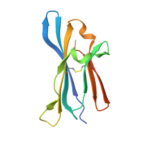
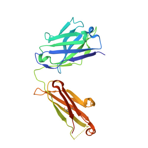
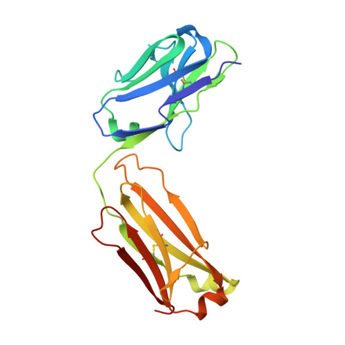
    7S7I

  • PubMed Abstract: 

    Characterization of antibody binding epitopes is an important factor in therapeutic drug discovery, as the binding site determines and drives antibody pharmacology and pharmacokinetics. Here, we present a novel application of carbene chemical footprinting with mass spectrometry for identification of antibody binding epitopes at the single-residue level. Two different photoactivated diazirine reagents provide complementary labeling information allowing structural refinement of the antibody binding interface. We applied this technique to map the epitopes of multiple MICA and CTLA-4 antibodies and validated the findings with X-ray crystallography and yeast surface display epitope mapping. The characterized epitopes were used to understand biolayer interferometry-derived competitive binding results at the structural level. We show that carbene footprinting provides fast and high-resolution epitope information critical in the antibody selection process and enables mechanistic understanding of function to accelerate the drug discovery process.


  • Organizational Affiliation

    Discovery Biotherapeutics, Bristol Myers Squibb, 700 Bay Road, Redwood City, California 94063, United States.


Macromolecules
Find similar proteins by:  (by identity cutoff)  |  3D Structure
Entity ID: 1
MoleculeChains Sequence LengthOrganismDetailsImage
MHC class I chain-related protein A103Homo sapiensMutation(s): 0 
Gene Names: MICA
UniProt
Find proteins for H9CTV0 (Homo sapiens)
Explore H9CTV0 
Go to UniProtKB:  H9CTV0
Entity Groups  
Sequence Clusters30% Identity50% Identity70% Identity90% Identity95% Identity100% Identity
UniProt GroupH9CTV0
Glycosylation
Glycosylation Sites: 1
Sequence Annotations
Expand
  • Reference Sequence
Find similar proteins by:  (by identity cutoff)  |  3D Structure
Entity ID: 2
MoleculeChains Sequence LengthOrganismDetailsImage
Fab heavy chain230Homo sapiensMutation(s): 0 
Entity Groups  
Sequence Clusters30% Identity50% Identity70% Identity90% Identity95% Identity100% Identity
Sequence Annotations
Expand
  • Reference Sequence
Find similar proteins by:  (by identity cutoff)  |  3D Structure
Entity ID: 3
MoleculeChains Sequence LengthOrganismDetailsImage
Fab light chain214Homo sapiensMutation(s): 0 
Entity Groups  
Sequence Clusters30% Identity50% Identity70% Identity90% Identity95% Identity100% Identity
Sequence Annotations
Expand
  • Reference Sequence
Experimental Data & Validation

Experimental Data

  • Method: X-RAY DIFFRACTION
  • Resolution: 2.40 Å
  • R-Value Free: 0.260 
  • R-Value Work: 0.220 
  • R-Value Observed: 0.222 
  • Space Group: P 1 21 1
Unit Cell:
Length ( Å )Angle ( ˚ )
a = 92.778α = 90
b = 170.432β = 105.81
c = 148.894γ = 90
Software Package:
Software NamePurpose
PHENIXrefinement
XDSdata reduction
XSCALEdata scaling
PHASERphasing

Structure Validation

View Full Validation Report


Ligand Structure Quality Assessment 


Entry History & Funding Information

Deposition Data


Funding OrganizationLocationGrant Number
Not funded--

Revision History  (Full details and data files)

  • Version 1.0: 2022-10-05
    Type: Initial release
  • Version 1.1: 2023-03-01
    Changes: Database references
  • Version 1.2: 2023-03-15
    Changes: Database references
  • Version 1.3: 2023-10-25
    Changes: Data collection, Refinement description