7S5J

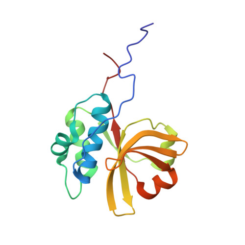
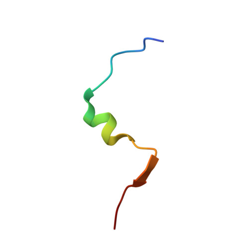
Solution NMR structure of substrate bound peptidase domain from PCAT1


Experimental Data Snapshot

  • Method: SOLUTION NMR
  • Conformers Calculated: 500 
  • Conformers Submitted: 20 
  • Selection Criteria: structures with the lowest energy 

wwPDB Validation   3D Report Full Report


This is version 1.2 of the entry. See complete history


Literature

Structural and dynamic studies of the peptidase domain from Clostridium thermocellum PCAT1.

Bhattacharya, S.Palillo, A.

(2022) Protein Sci 31: 498-512

  • DOI: https://doi.org/10.1002/pro.4248
  • Primary Citation of Related Structures:  
    7N87, 7S5J

  • PubMed Abstract: 

    The export of antimicrobial peptides is mediated by diverse mechanisms in bacterial quorum sensing pathways. One such binary system employed by gram-positive bacteria is the PCAT1 ABC transporter coupled to a cysteine protease. The focus of this study is the N-terminal C39 peptidase (PEP) domain from Clostridium thermocellum PCAT1 that processes its natural substrate CtA by cleaving a conserved -GG- motif to separate the cargo from the leader peptide prior to secretion. In this study, we are primarily interested in elucidating the dynamic and structural determinants of CtA binding and how it is coupled to cleavage efficiency in the PCAT1 PEP domain. To this end, we have characterized CtA interactions with PEP domain and PCAT1 transporter in detergent micelles using solution nuclear magnetic resonance spectroscopy. The bound CtA structure revealed the disordered C-terminal cargo peptide is linked by a sterically hindered cleavage site to a helix docked within a hydrophobic cavity in the PEP domain. The wide range of internal motions detected by amide nitrogen (N 15 ) relaxation measurements in the free enzyme and substrate-bound complex suggests the binding site is relatively floppy. This flexibility plays a key role in the structural rearrangement necessary to relax steric inhibition in the bound substrate. In conjunction with previously reported PCAT1 structures, we offer fresh insight into the ATP-mediated association between PEP and transmembrane domains as a putative mechanism to optimize peptide cleavage by regulating the width and flexibility of the enzyme active site.


  • Organizational Affiliation

    New York Structural Biology Center, New York, New York, USA.


Macromolecules
Find similar proteins by:  (by identity cutoff)  |  3D Structure
Entity ID: 1
MoleculeChains Sequence LengthOrganismDetailsImage
Peptidase C39151Acetivibrio thermocellus ATCC 27405Mutation(s): 1 
Gene Names: Cthe_0534
UniProt
Find proteins for A3DCU1 (Acetivibrio thermocellus (strain ATCC 27405 / DSM 1237 / JCM 9322 / NBRC 103400 / NCIMB 10682 / NRRL B-4536 / VPI 7372))
Explore A3DCU1 
Go to UniProtKB:  A3DCU1
Entity Groups  
Sequence Clusters30% Identity50% Identity70% Identity90% Identity95% Identity100% Identity
UniProt GroupA3DCU1
Sequence Annotations
Expand
  • Reference Sequence

Find similar proteins by:  Sequence   |   3D Structure  

Entity ID: 2
MoleculeChains Sequence LengthOrganismDetailsImage
CtA peptide24Acetivibrio thermocellus ATCC 27405Mutation(s): 0 
Gene Names: Cthe_0535
UniProt
Find proteins for A3DCU2 (Acetivibrio thermocellus (strain ATCC 27405 / DSM 1237 / JCM 9322 / NBRC 103400 / NCIMB 10682 / NRRL B-4536 / VPI 7372))
Explore A3DCU2 
Go to UniProtKB:  A3DCU2
Entity Groups  
Sequence Clusters30% Identity50% Identity70% Identity90% Identity95% Identity100% Identity
UniProt GroupA3DCU2
Sequence Annotations
Expand
  • Reference Sequence
Experimental Data & Validation

Experimental Data

  • Method: SOLUTION NMR
  • Conformers Calculated: 500 
  • Conformers Submitted: 20 
  • Selection Criteria: structures with the lowest energy 

Structure Validation

View Full Validation Report


Entry History & Funding Information

Deposition Data


Funding OrganizationLocationGrant Number
National Institutes of Health/National Cancer Institute (NIH/NCI)United States1P41GM118302-01A1

Revision History  (Full details and data files)

  • Version 1.0: 2021-12-29
    Type: Initial release
  • Version 1.1: 2022-02-23
    Changes: Database references
  • Version 1.2: 2024-05-15
    Changes: Data collection, Database references