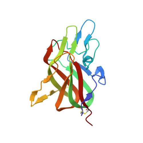
7S0P

Crystal structure of Porcine Factor VIII C2 Domain Bound to Phosphatidylserine


Experimental Data Snapshot

  • Method: X-RAY DIFFRACTION
  • Resolution: 1.30 Å
  • R-Value Free: 0.156 
  • R-Value Work: 0.137 
  • R-Value Observed: 0.137 

Starting Model: experimental
View more details

wwPDB Validation   3D Report Full Report


Ligand Structure Quality Assessment 


This is version 1.4 of the entry. See complete history


Literature

Stable binding to phosphatidylserine-containing membranes requires conserved arginine residues in tandem C domains of blood coagulation factor VIII.

Peters, S.C.Childers, K.C.Mitchell, C.E.Avery, N.G.Reese Jr., S.S.Mitchell, C.Wo, S.W.Swanson, C.D.Brison, C.M.Spiegel Jr., P.C.

(2022) Front Mol Biosci 9: 1040106-1040106

  • DOI: https://doi.org/10.3389/fmolb.2022.1040106
  • Primary Citation of Related Structures:  
    7S0P

  • PubMed Abstract: 

    At sites of vascular damage, factor VIII (fVIII) is proteolytically activated by thrombin and binds to activated platelet surfaces with activated factor IX (fIXa) to form the intrinsic "tenase" complex. Previous structural and mutational studies of fVIII have identified the C1 and C2 domains in binding to negatively charged membrane surfaces through β-hairpin loops with solvent-exposed hydrophobic residues and a ring of positively charged basic residues. Several hemophilia A-associated mutations within the C domains are suggested to disrupt lipid binding, preventing formation of the intrinsic tenase complex. In this study, we devised a novel platform for generating recombinant C1, C2, and C1C2 domain constructs and performed mutagenesis of several charged residues proximal to the putative membrane binding region of each C domain. Binding measurements between phosphatidylserine (PS)-containing lipid membrane surfaces and fVIII C domains demonstrated an ionic strength dependence on membrane binding affinity. Mutations to basic residues adjacent to the surface-exposed hydrophobic regions of C1 and C2 differentially disrupted membrane binding, with abrogation of binding occurring for mutations to conserved arginine residues in the C1 (R2163) and C2 (R2320) domains. Lastly, we determined the X-ray crystal structure of the porcine fVIII C2 domain bound to o -phospho-L-serine, the polar headgroup of PS, which binds to a basic cleft and makes charge-charge contact with R2320. We conclude that basic clefts in the fVIII C domains bind to PS-containing membranes through conserved arginine residues via a C domain modularity, where each C domain possesses modest electrostatic-dependent affinity and tandem C domains are required for high affinity binding.


  • Organizational Affiliation

    Department of Chemistry, Western Washington University, Bellingham, WA, United States.


Macromolecules
Find similar proteins by:  (by identity cutoff)  |  3D Structure
Entity ID: 1
MoleculeChains Sequence LengthOrganismDetailsImage
Coagulation factor VIIIA [auth M]157Sus scrofaMutation(s): 0 
Gene Names: F8CF8
UniProt
Find proteins for P12263 (Sus scrofa)
Explore P12263 
Go to UniProtKB:  P12263
Entity Groups  
Sequence Clusters30% Identity50% Identity70% Identity90% Identity95% Identity100% Identity
UniProt GroupP12263
Sequence Annotations
Expand
  • Reference Sequence
Small Molecules
Ligands 1 Unique
IDChains Name / Formula / InChI Key2D Diagram3D Interactions
SEP (Subject of Investigation/LOI)
Query on SEP

Download Ideal Coordinates CCD File 
B [auth M]PHOSPHOSERINE
C3 H8 N O6 P
BZQFBWGGLXLEPQ-REOHCLBHSA-N
Experimental Data & Validation

Experimental Data

  • Method: X-RAY DIFFRACTION
  • Resolution: 1.30 Å
  • R-Value Free: 0.156 
  • R-Value Work: 0.137 
  • R-Value Observed: 0.137 
  • Space Group: I 2 2 2
Unit Cell:
Length ( Å )Angle ( ˚ )
a = 49.072α = 90
b = 68.323β = 90
c = 105.987γ = 90
Software Package:
Software NamePurpose
PHENIXrefinement
Adxvdata reduction
Aimlessdata scaling
PHASERphasing
Cootmodel building
XDSdata reduction

Structure Validation

View Full Validation Report


Ligand Structure Quality Assessment 


Entry History & Funding Information

Deposition Data


Funding OrganizationLocationGrant Number
National Science Foundation (NSF, United States)United StatesMRI 1429164
National Institutes of Health/National Heart, Lung, and Blood Institute (NIH/NHLBI)United StatesR15HL103518
National Institutes of Health/National Heart, Lung, and Blood Institute (NIH/NHLBI)United StatesU54HL141981
National Institutes of Health/National Heart, Lung, and Blood Institute (NIH/NHLBI)United StatesR44HL117511
National Institutes of Health/National Heart, Lung, and Blood Institute (NIH/NHLBI)United StatesR44HL110448
National Institutes of Health/National Heart, Lung, and Blood Institute (NIH/NHLBI)United StatesU54HL112309

Revision History  (Full details and data files)

  • Version 1.0: 2021-10-27
    Type: Initial release
  • Version 1.1: 2022-11-09
    Changes: Database references
  • Version 1.2: 2022-11-30
    Changes: Database references
  • Version 1.3: 2023-10-25
    Changes: Data collection, Refinement description
  • Version 1.4: 2024-10-30
    Changes: Structure summary