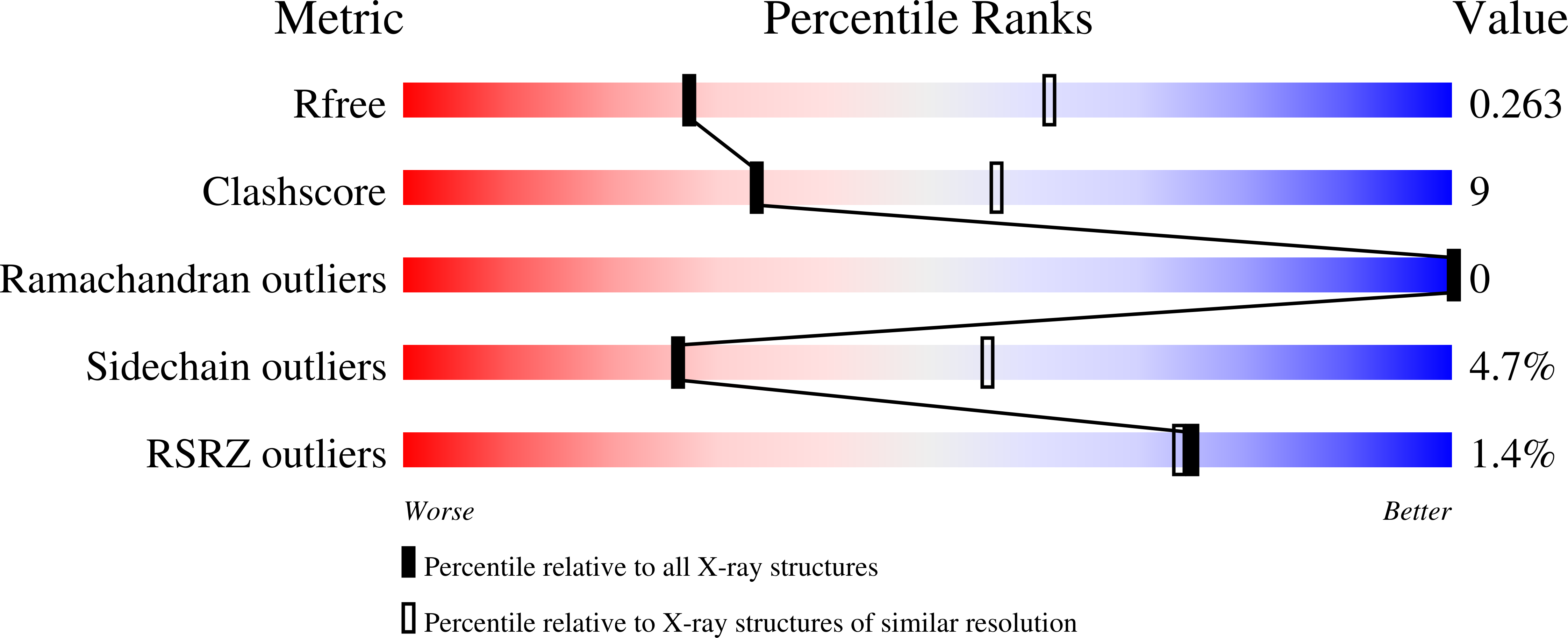
7LRN

Structure of the Siderophore Interacting Protein from Acinetbacter baumannii


Experimental Data Snapshot

  • Method: X-RAY DIFFRACTION
  • Resolution: 2.85 Å
  • R-Value Free: 0.264 
  • R-Value Work: 0.218 
  • R-Value Observed: 0.221 

Starting Model: experimental
View more details

wwPDB Validation   3D Report Full Report


Ligand Structure Quality Assessment 


This is version 1.2 of the entry. See complete history


Literature

Structural and Biochemical Characterization of the Flavin-Dependent Siderophore-Interacting Protein from Acinetobacter baumannii .

Valentino, H.Korasick, D.A.Bohac, T.J.Shapiro, J.A.Wencewicz, T.A.Tanner, J.J.Sobrado, P.

(2021) ACS Omega 6: 18537-18547

  • DOI: https://doi.org/10.1021/acsomega.1c03047
  • Primary Citation of Related Structures:  
    7LRN

  • PubMed Abstract: 

    Acinetobacter baumannii is an opportunistic pathogen with a high mortality rate due to multi-drug-resistant strains. The synthesis and uptake of the iron-chelating siderophores acinetobactin (Acb) and preacinetobactin (pre-Acb) have been shown to be essential for virulence. Here, we report the kinetic and structural characterization of BauF, a flavin-dependent siderophore-interacting protein (SIP) required for the reduction of Fe(III) bound to Acb/pre-Acb and release of Fe(II). Stopped-flow spectrophotometric studies of the reductive half-reaction show that BauF forms a stable neutral flavin semiquinone intermediate. Reduction with NAD(P)H is very slow ( k obs , 0.001 s -1 ) and commensurate with the rate of reduction by photobleaching, suggesting that NAD(P)H are not the physiological partners of BauF. The reduced BauF was oxidized by Acb-Fe ( k obs , 0.02 s -1 ) and oxazole pre-Acb-Fe (ox-pre-Acb-Fe) ( k obs , 0.08 s -1 ), a rigid analogue of pre-Acb, at a rate 3-11 times faster than that with molecular oxygen alone. The structure of FAD-bound BauF was solved at 2.85 Å and was found to share a similarity to Shewanella SIPs. The biochemical and structural data presented here validate the role of BauF in A. baumannii iron assimilation and provide information important for drug design.


  • Organizational Affiliation

    Department of Biochemistry, Virginia Tech, Blacksburg, Virginia 24061, United States.


Macromolecules
Find similar proteins by:  (by identity cutoff)  |  3D Structure
Entity ID: 1
MoleculeChains Sequence LengthOrganismDetailsImage
NADPH-dependent ferric siderophore reductase
A, B
301Acinetobacter baumanniiMutation(s): 0 
Gene Names: A7M79_07860BGC29_02895
UniProt
Find proteins for A0A1E3M8P2 (Acinetobacter baumannii)
Explore A0A1E3M8P2 
Go to UniProtKB:  A0A1E3M8P2
Entity Groups  
Sequence Clusters30% Identity50% Identity70% Identity90% Identity95% Identity100% Identity
UniProt GroupA0A1E3M8P2
Sequence Annotations
Expand
  • Reference Sequence
Experimental Data & Validation

Experimental Data

  • Method: X-RAY DIFFRACTION
  • Resolution: 2.85 Å
  • R-Value Free: 0.264 
  • R-Value Work: 0.218 
  • R-Value Observed: 0.221 
  • Space Group: C 2 2 21
Unit Cell:
Length ( Å )Angle ( ˚ )
a = 86.79α = 90
b = 127.252β = 90
c = 150.304γ = 90
Software Package:
Software NamePurpose
Aimlessdata scaling
PHENIXrefinement
PDB_EXTRACTdata extraction
XDSdata reduction
BALBESphasing

Structure Validation

View Full Validation Report


Ligand Structure Quality Assessment 


Entry History & Funding Information

Deposition Data


Funding OrganizationLocationGrant Number
National Science Foundation (NSF, United States)United StatesCHE-2003986

Revision History  (Full details and data files)

  • Version 1.0: 2021-07-21
    Type: Initial release
  • Version 1.1: 2021-08-25
    Changes: Database references
  • Version 1.2: 2023-10-18
    Changes: Data collection, Refinement description