7LPG

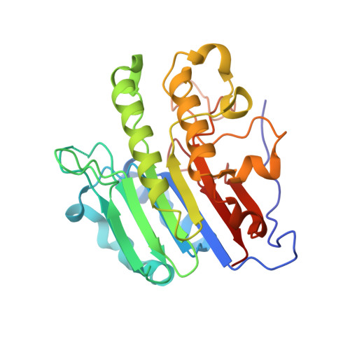
APE1 product complex with abasic ribonucleotide DNA


Experimental Data Snapshot

  • Method: X-RAY DIFFRACTION
  • Resolution: 2.08 Å
  • R-Value Free: 0.263 
  • R-Value Work: 0.219 
  • R-Value Observed: 0.221 

Starting Model: experimental
View more details

wwPDB Validation   3D Report Full Report


This is version 1.1 of the entry. See complete history


Literature

Altered APE1 activity on abasic ribonucleotides is mediated by changes in the nucleoside sugar pucker.

Hoitsma, N.M.Click, T.H.Agarwal, P.K.Freudenthal, B.D.

(2021) Comput Struct Biotechnol J 19: 3293-3302

  • DOI: https://doi.org/10.1016/j.csbj.2021.05.035
  • Primary Citation of Related Structures:  
    7LPG, 7LPH, 7LPI, 7LPJ

  • PubMed Abstract: 

    Ribonucleotides (rNTPs) are predicted to be incorporated into the genome at a rate of up to 3 million times per cell division, making rNTPs the most common non-standard nucleotide in the human genome. Typically, misinserted ribonucleotides are repaired by the ribonucleotide excision repair (RER) pathway, which is initiated by RNase H2 cleavage. However, rNTPs are susceptible to spontaneous depurination generating abasic ribonucleotides (rAPs), which are unable to be processed by RNase H2. Additionally, rAPs have been found in nascent RNA and coupled to R-loops. Recent work identified that base excision repair (BER) protein AP-Endonuclease 1 (APE1) is responsible for the initial processing of rAPs embedded in DNA and in R-loops. APE1 is a well characterized AP endonuclease that cleaves 5' of abasic sites, but its ability to cleave at rAPs remains poorly understood. Here, we utilize enzyme kinetics, X-ray crystallography, and molecular dynamics simulations to provide insight into rAP processing by APE1. Enzyme kinetics were used to determine pre-steady-state rates of APE1 cleavage on DNA substrates containing rAP, revealing a decrease in activity compared to cleavage at a canonical deoxy-AP substrate. Using X-ray crystallography, we identified novel contacts between the rAP and the APE1 active site. We demonstrate that the rAP sugar pucker is accommodated in the active site in a C3'-endo conformation, influencing its position and contributing to a decrease in activity compared to the deoxy-AP site. Together, this work provides molecular level insights into rAP processing by APE1 and advances our understanding of ribonucleotide processing within genomic DNA.


  • Organizational Affiliation

    Department of Biochemistry and Molecular Biology, University of Kansas Medical Center, Kansas City, KS 66160, USA.


Macromolecules

Find similar proteins by:  (by identity cutoff)  |  3D Structure
Entity ID: 1
MoleculeChains Sequence LengthOrganismDetailsImage
DNA-(apurinic or apyrimidinic site) lyaseA [auth B],
C [auth D]
276Homo sapiensMutation(s): 1 
Gene Names: APEX1APEAPE1APEXAPXHAP1REF1
EC: 3.1 (PDB Primary Data), 4.2.99.18 (PDB Primary Data), 3.1.21 (UniProt), 3.1.11.2 (UniProt)
UniProt & NIH Common Fund Data Resources
Find proteins for P27695 (Homo sapiens)
Explore P27695 
Go to UniProtKB:  P27695
PHAROS:  P27695
GTEx:  ENSG00000100823 
Entity Groups  
Sequence Clusters30% Identity50% Identity70% Identity90% Identity95% Identity100% Identity
UniProt GroupP27695
Sequence Annotations
Expand
  • Reference Sequence

Find similar nucleic acids by:  Sequence   |   3D Structure  

Entity ID: 2
MoleculeChains LengthOrganismImage
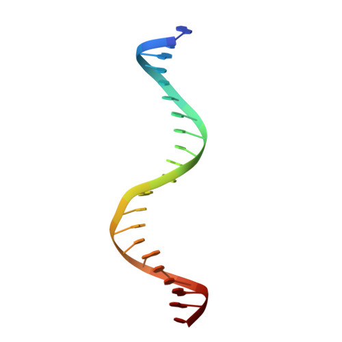
DNA (5'-D(*GP*GP*AP*TP*CP*CP*GP*TP*CP*GP*AP*GP*CP*GP*CP*AP*TP*CP*AP*GP*C)-3')B [auth C]21synthetic construct
Sequence Annotations
Expand
  • Reference Sequence

Find similar nucleic acids by:  Sequence   |   3D Structure  

Entity ID: 3
MoleculeChains LengthOrganismImage
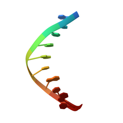
DNA (5'-D(*GP*CP*TP*GP*AP*TP*GP*CP*GP*C)-3')D [auth E]10synthetic construct
Sequence Annotations
Expand
  • Reference Sequence

Find similar nucleic acids by:  Sequence   |   3D Structure  

Entity ID: 4
MoleculeChains LengthOrganismImage
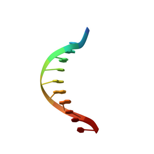
DNA (5'-R(P*N)-D(P*CP*GP*AP*CP*GP*GP*AP*TP*CP*C)-3')E [auth A]11synthetic construct
Sequence Annotations
Expand
  • Reference Sequence
Experimental Data & Validation

Experimental Data

  • Method: X-RAY DIFFRACTION
  • Resolution: 2.08 Å
  • R-Value Free: 0.263 
  • R-Value Work: 0.219 
  • R-Value Observed: 0.221 
  • Space Group: P 1
Unit Cell:
Length ( Å )Angle ( ˚ )
a = 44.118α = 83.682
b = 61.077β = 78.066
c = 73.039γ = 87.491
Software Package:
Software NamePurpose
PHENIXrefinement
HKL-2000data collection
HKL-2000data scaling
PDB_EXTRACTdata extraction
HKL-2000data reduction
PHENIXphasing

Structure Validation

View Full Validation Report


Entry History & Funding Information

Deposition Data


Funding OrganizationLocationGrant Number
National Institutes of Health/National Institute of Environmental Health Sciences (NIH/NIEHS)United StatesR01 ES029203

Revision History  (Full details and data files)

  • Version 1.0: 2021-08-04
    Type: Initial release
  • Version 1.1: 2023-10-18
    Changes: Data collection, Database references, Refinement description