7JLG

Human PrimPol extending from the erroneous primer base A opposite the 8-oxoguanine lesion


Experimental Data Snapshot

  • Method: X-RAY DIFFRACTION
  • Resolution: 2.07 Å
  • R-Value Free: 0.219 
  • R-Value Work: 0.201 
  • R-Value Observed: 0.202 

Starting Model: experimental
View more details

wwPDB Validation   3D Report Full Report


Ligand Structure Quality Assessment 


This is version 1.2 of the entry. See complete history


Literature

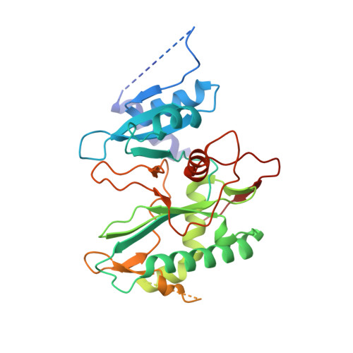
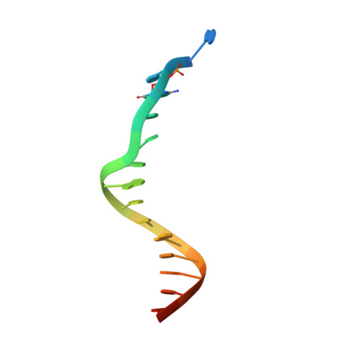
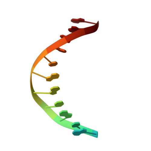
Structural basis of DNA synthesis opposite 8-oxoguanine by human PrimPol primase-polymerase.

Rechkoblit, O.Johnson, R.E.Gupta, Y.K.Prakash, L.Prakash, S.Aggarwal, A.K.

(2021) Nat Commun 12: 4020-4020

  • DOI: https://doi.org/10.1038/s41467-021-24317-z
  • Primary Citation of Related Structures:  
    7JK1, 7JKL, 7JKP, 7JL8, 7JLG

  • PubMed Abstract: 

    PrimPol is a human DNA polymerase-primase that localizes to mitochondria and nucleus and bypasses the major oxidative lesion 7,8-dihydro-8-oxoguanine (oxoG) via translesion synthesis, in mostly error-free manner. We present structures of PrimPol insertion complexes with a DNA template-primer and correct dCTP or erroneous dATP opposite the lesion, as well as extension complexes with C or A as a 3'-terminal primer base. We show that during the insertion of C and extension from it, the active site is unperturbed, reflecting the readiness of PrimPol to accommodate oxoG(anti). The misinsertion of A opposite oxoG(syn) also does not alter the active site, and is likely less favorable due to lower thermodynamic stability of the oxoG(syn)•A base-pair. During the extension step, oxoG(syn) induces an opening of its base-pair with A or misalignment of the 3'-A primer terminus. Together, the structures show how PrimPol accurately synthesizes DNA opposite oxidatively damaged DNA in human cells.


  • Organizational Affiliation

    Department of Pharmacological Sciences, Icahn School of Medicine at Mount Sinai, New York, NY, USA. olga.rechkoblit@mssm.edu.


Macromolecules

Find similar proteins by:  (by identity cutoff)  |  3D Structure
Entity ID: 1
MoleculeChains Sequence LengthOrganismDetailsImage
DNA-directed primase/polymerase proteinA,
D [auth B]
354Homo sapiensMutation(s): 0 
Gene Names: PRIMPOLCCDC111
EC: 2.7.7
UniProt & NIH Common Fund Data Resources
Find proteins for Q96LW4 (Homo sapiens)
Explore Q96LW4 
Go to UniProtKB:  Q96LW4
PHAROS:  Q96LW4
GTEx:  ENSG00000164306 
Entity Groups  
Sequence Clusters30% Identity50% Identity70% Identity90% Identity95% Identity100% Identity
UniProt GroupQ96LW4
Sequence Annotations
Expand
  • Reference Sequence

Find similar nucleic acids by:  Sequence   |   3D Structure  

Entity ID: 2
MoleculeChains LengthOrganismImage
DNA (5'-D(P*AP*TP*(8OG)P*CP*CP*TP*AP*CP*CP*AP*CP*AP*CP*CP*C)-3')B [auth C],
E [auth G]
17synthetic construct
Sequence Annotations
Expand
  • Reference Sequence

Find similar nucleic acids by:  Sequence   |   3D Structure  

Entity ID: 3
MoleculeChains LengthOrganismImage
DNA (5'-D(P*GP*TP*GP*TP*GP*GP*TP*AP*GP*GP*A)-3')C [auth D],
F [auth H]
13synthetic construct
Sequence Annotations
Expand
  • Reference Sequence
Small Molecules
Ligands 4 Unique
IDChains Name / Formula / InChI Key2D Diagram3D Interactions
DTP
Query on DTP

Download Ideal Coordinates CCD File 
G [auth A],
J [auth B]
2'-DEOXYADENOSINE 5'-TRIPHOSPHATE
C10 H16 N5 O12 P3
SUYVUBYJARFZHO-RRKCRQDMSA-N
PEG
Query on PEG

Download Ideal Coordinates CCD File 
M [auth B]DI(HYDROXYETHYL)ETHER
C4 H10 O3
MTHSVFCYNBDYFN-UHFFFAOYSA-N
GOL
Query on GOL

Download Ideal Coordinates CCD File 
N [auth B],
O [auth B]
GLYCEROL
C3 H8 O3
PEDCQBHIVMGVHV-UHFFFAOYSA-N
CA
Query on CA

Download Ideal Coordinates CCD File 
H [auth A],
I [auth A],
K [auth B],
L [auth B]
CALCIUM ION
Ca
BHPQYMZQTOCNFJ-UHFFFAOYSA-N
Experimental Data & Validation

Experimental Data

  • Method: X-RAY DIFFRACTION
  • Resolution: 2.07 Å
  • R-Value Free: 0.219 
  • R-Value Work: 0.201 
  • R-Value Observed: 0.202 
  • Space Group: P 1
Unit Cell:
Length ( Å )Angle ( ˚ )
a = 51.031α = 71.36
b = 65.21β = 84.31
c = 73.115γ = 88.63
Software Package:
Software NamePurpose
PHENIXrefinement
PDB_EXTRACTdata extraction
XDSdata reduction
XDSdata scaling
PHASERphasing

Structure Validation

View Full Validation Report


Ligand Structure Quality Assessment 


Entry History & Funding Information

Deposition Data


Funding OrganizationLocationGrant Number
National Institutes of Health/National Institute of General Medical Sciences (NIH/NIGMS)United StatesR35-GM131780

Revision History  (Full details and data files)

  • Version 1.0: 2021-06-30
    Type: Initial release
  • Version 1.1: 2021-07-14
    Changes: Database references
  • Version 1.2: 2023-10-18
    Changes: Data collection, Database references, Refinement description