7E5B

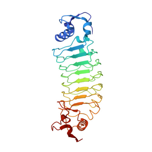
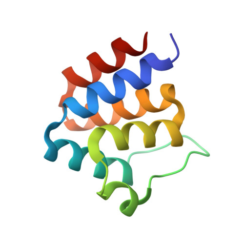
Crystal structure of ASC PYD Domain and Rb-B7


Experimental Data Snapshot

  • Method: X-RAY DIFFRACTION
  • Resolution: 2.29 Å
  • R-Value Free: 0.236 
  • R-Value Work: 0.195 
  • R-Value Observed: 0.197 

Starting Model: experimental
View more details

wwPDB Validation   3D Report Full Report


This is version 1.2 of the entry. See complete history


Literature

Oligomeric states of ASC specks regulate inflammatory responses by inflammasome in the extracellular space.

Yu, T.G.Cha, J.S.Kim, G.Sohn, Y.K.Yoo, Y.Kim, U.Song, J.J.Cho, H.S.Kim, H.S.

(2023) Cell Death Discov 9: 142-142

  • DOI: https://doi.org/10.1038/s41420-023-01438-6
  • Primary Citation of Related Structures:  
    7E5B

  • PubMed Abstract: 

    Inflammasomes are multi-protein complexes and play a crucial role in host defense against pathogens. Downstream inflammatory responses through inflammasomes are known to be related to the oligomerization degree of ASC specks, but the detailed mechanism still remains unexplored. Here, we demonstrate that oligomerization degrees of ASC specks regulate the caspase-1 activation in the extracellular space. A protein binder specific for a pyrin domain (PYD) of ASC (ASC PYD ) was developed, and structural analysis revealed that the protein binder effectively inhibits the interaction between PYDs, disassembling ASC specks into low oligomeric states. ASC specks with a low oligomerization degree were shown to enhance the activation of caspase-1 by recruiting and processing more premature caspase-1 through interactions between CARD of caspase-1 (caspase-1 CARD ) and CARD of ASC (ASC CARD ). These findings can provide insight into controlling the inflammasome-mediated inflammatory process as well as the development of inflammasome-targeting drugs.


  • Organizational Affiliation

    Departement of Biological Sciences, Korea Advanced Institute of Science and Technology (KAIST), Daejeon, 34141, Korea.


Macromolecules
Find similar proteins by:  (by identity cutoff)  |  3D Structure
Entity ID: 1
MoleculeChains Sequence LengthOrganismDetailsImage
Repebody (Rb-B7)
A, B
266CyclostomataMutation(s): 0 
Entity Groups  
Sequence Clusters30% Identity50% Identity70% Identity90% Identity95% Identity100% Identity
Sequence Annotations
Expand
  • Reference Sequence
Find similar proteins by:  (by identity cutoff)  |  3D Structure
Entity ID: 2
MoleculeChains Sequence LengthOrganismDetailsImage
Apoptosis-associated speck-like protein containing a CARD
C, D
92Homo sapiensMutation(s): 0 
Gene Names: PYCARDASCCARD5TMS1
UniProt & NIH Common Fund Data Resources
Find proteins for Q9ULZ3 (Homo sapiens)
Explore Q9ULZ3 
Go to UniProtKB:  Q9ULZ3
PHAROS:  Q9ULZ3
GTEx:  ENSG00000103490 
Entity Groups  
Sequence Clusters30% Identity50% Identity70% Identity90% Identity95% Identity100% Identity
UniProt GroupQ9ULZ3
Sequence Annotations
Expand
  • Reference Sequence
Experimental Data & Validation

Experimental Data

  • Method: X-RAY DIFFRACTION
  • Resolution: 2.29 Å
  • R-Value Free: 0.236 
  • R-Value Work: 0.195 
  • R-Value Observed: 0.197 
  • Space Group: P 43 21 2
Unit Cell:
Length ( Å )Angle ( ˚ )
a = 79.481α = 90
b = 79.481β = 90
c = 279.197γ = 90
Software Package:
Software NamePurpose
PHENIXrefinement
HKL-2000data reduction
HKL-2000data scaling
PHENIXphasing

Structure Validation

View Full Validation Report


Entry History 

Deposition Data

Revision History  (Full details and data files)

  • Version 1.0: 2022-03-02
    Type: Initial release
  • Version 1.1: 2023-05-10
    Changes: Database references
  • Version 1.2: 2023-11-29
    Changes: Data collection, Refinement description