7E48

Crystal structure of InhA in complex with 3-nitropropanoic acid inhibitor


Experimental Data Snapshot

  • Method: X-RAY DIFFRACTION
  • Resolution: 2.50 Å
  • R-Value Free: 0.220 
  • R-Value Work: 0.185 

Starting Model: experimental
View more details

wwPDB Validation   3D Report Full Report


Ligand Structure Quality Assessment 


This is version 1.2 of the entry. See complete history


Literature

Inhibition of Mycobacterium tuberculosis InhA by 3-nitropropanoic acid.

Songsiriritthigul, C.Hanwarinroj, C.Pakamwong, B.Srimanote, P.Suttipanta, N.Sureram, S.Suttisintong, K.Kamsri, P.Punkvang, A.Spencer, J.Kittakoop, P.Pungpo, P.

(2022) Proteins 90: 898-904

  • DOI: https://doi.org/10.1002/prot.26268
  • Primary Citation of Related Structures:  
    7E48

  • PubMed Abstract: 

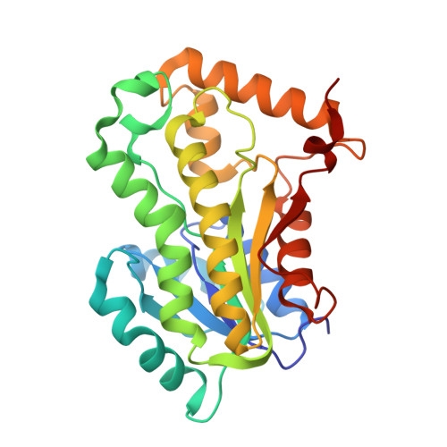
    3-Nitropropanoic acid (3NP), a bioactive fungal natural product, was previously demonstrated to inhibit growth of Mycobacterium tuberculosis. Here we demonstrate that 3NP inhibits the 2-trans-enoyl-acyl carrier protein reductase (InhA) from Mycobacterium tuberculosis with an IC 50 value of 71 μM, and present the crystal structure of the ternary InhA-NAD + -3NP complex. The complex contains the InhA substrate-binding loop in an ordered, open conformation with Tyr158, a catalytically important residue whose orientation defines different InhA substrate/inhibitor complex conformations, in the "out" position. 3NP occupies a hydrophobic binding site adjacent to the NAD + cofactor and close to that utilized by the diphenyl ether triclosan, but binds predominantly via electrostatic and water-mediated hydrogen-bonding interactions with the protein backbone and NAD + cofactor. The identified mode of 3NP binding provides opportunities to improve inhibitory activity toward InhA.


  • Organizational Affiliation

    Synchrotron Light Research Institute (Public Organization), Nakhon Ratchasima, Thailand.


Macromolecules
Find similar proteins by:  (by identity cutoff)  |  3D Structure
Entity ID: 1
MoleculeChains Sequence LengthOrganismDetailsImage
Enoyl-[acyl-carrier-protein] reductase [NADH]
A, B, C, D
269Mycobacterium tuberculosis H37RvMutation(s): 0 
Gene Names: inhARv1484MTCY277.05
EC: 1.3.1.9
UniProt
Find proteins for P9WGR1 (Mycobacterium tuberculosis (strain ATCC 25618 / H37Rv))
Explore P9WGR1 
Go to UniProtKB:  P9WGR1
Entity Groups  
Sequence Clusters30% Identity50% Identity70% Identity90% Identity95% Identity100% Identity
UniProt GroupP9WGR1
Sequence Annotations
Expand
  • Reference Sequence
Small Molecules
Ligands 3 Unique
IDChains Name / Formula / InChI Key2D Diagram3D Interactions
NAD
Query on NAD

Download Ideal Coordinates CCD File 
E [auth A],
G [auth B],
J [auth C],
L [auth D]
NICOTINAMIDE-ADENINE-DINUCLEOTIDE
C21 H27 N7 O14 P2
BAWFJGJZGIEFAR-NNYOXOHSSA-N
3NP
Query on 3NP

Download Ideal Coordinates CCD File 
F [auth A],
H [auth B],
K [auth C],
M [auth D]
3-NITROPROPANOIC ACID
C3 H5 N O4
WBLZUCOIBUDNBV-UHFFFAOYSA-N
GOL
Query on GOL

Download Ideal Coordinates CCD File 
I [auth B]GLYCEROL
C3 H8 O3
PEDCQBHIVMGVHV-UHFFFAOYSA-N
Experimental Data & Validation

Experimental Data

  • Method: X-RAY DIFFRACTION
  • Resolution: 2.50 Å
  • R-Value Free: 0.220 
  • R-Value Work: 0.185 
  • Space Group: P 32
Unit Cell:
Length ( Å )Angle ( ˚ )
a = 96.39α = 90
b = 96.39β = 90
c = 139.5γ = 120
Software Package:
Software NamePurpose
REFMACrefinement
MOSFLMdata reduction
SCALAdata scaling
PHASERphasing

Structure Validation

View Full Validation Report


Ligand Structure Quality Assessment 


Entry History 

Deposition Data

Revision History  (Full details and data files)

  • Version 1.0: 2021-11-24
    Type: Initial release
  • Version 1.1: 2022-02-23
    Changes: Database references
  • Version 1.2: 2023-11-29
    Changes: Data collection, Derived calculations, Refinement description