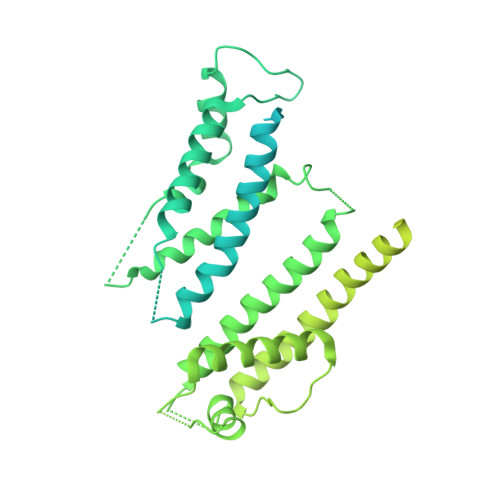
Cryo-EM Structure of K + -Bound hERG Channel Complexed with the Blocker Astemizole.
Asai, T., Adachi, N., Moriya, T., Oki, H., Maru, T., Kawasaki, M., Suzuki, K., Chen, S., Ishii, R., Yonemori, K., Igaki, S., Yasuda, S., Ogasawara, S., Senda, T., Murata, T.(2021) Structure 29: 203-212.e4
- PubMed: 33450182
- DOI: https://doi.org/10.1016/j.str.2020.12.007
- Primary Citation of Related Structures:
7CN0, 7CN1 - PubMed Abstract:
The hERG channel is a voltage-gated potassium channel involved in cardiac repolarization. Off-target hERG inhibition by drugs has become a critical issue in the pharmaceutical industry. The three-dimensional structure of the hERG channel was recently reported at 3.8-Å resolution using cryogenic electron microscopy (cryo-EM). However, the drug inhibition mechanism remains unclear because of the scarce structural information regarding the drug- and potassium-bound hERG channels. In this study, we obtained the cryo-EM density map of potassium-bound hERG channel complexed with astemizole, a well-known hERG inhibitor that increases risk of potentially fatal arrhythmia, at 3.5-Å resolution. The structure suggested that astemizole inhibits potassium conduction by binding directly below the selectivity filter. Furthermore, we propose a possible binding model of astemizole to the hERG channel and provide insights into the unusual sensitivity of hERG to several drugs.
Organizational Affiliation:
Department of Chemistry, Graduate School of Science, Chiba University, 1-33 Yayoi-cho, Inage, Chiba 263-8522, Japan.


