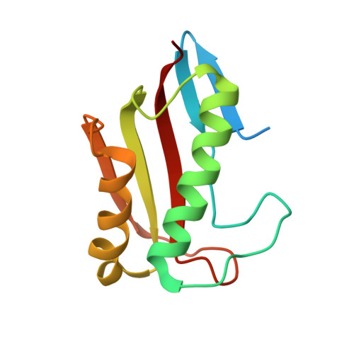
7CD3

Crystal structure of the S103F mutant of Bacillus subtilis (natto) YabJ protein.


Experimental Data Snapshot

  • Method: X-RAY DIFFRACTION
  • Resolution: 2.10 Å
  • R-Value Free: 0.253 
  • R-Value Work: 0.194 
  • R-Value Observed: 0.198 

Starting Model: experimental
View more details

wwPDB Validation   3D Report Full Report


This is version 1.1 of the entry. See complete history


Literature

Tetramer formation of Bacillus subtilis YabJ protein that belongs to YjgF/YER057c/UK114 family.

Fujimoto, Z.Hong, L.T.T.Kishine, N.Suzuki, N.Kimura, K.

(2021) Biosci Biotechnol Biochem 85: 297-306

  • DOI: https://doi.org/10.1093/bbb/zbaa037
  • Primary Citation of Related Structures:  
    5Y6U, 7CD2, 7CD3, 7CD4

  • PubMed Abstract: 

    Bacillus subtilis YabJ protein belongs to the highly conserved YjgF/YER057c/UK114 family, which has a homotrimeric quaternary structure. The dominant allele of yabJ gene that is caused by a single amino acid mutation of Ser103Phe enables poly-γ-glutamic acid (γPGA) production of B. subtilis under conditions where the cell-density signal transduction was disturbed by the loss of DegQ function. X-ray crystallography of recombinant proteins revealed that unlike the homotrimeric wild-type YabJ, the mutant YabJ(Ser103Phe) had a homotetrameric quaternary structure, and the structural change appeared to be triggered by an inversion of the fifth β-strand. The YabJ homotetramer has a hole that is highly accessible, penetrating through the tetramer, and 2 surface concaves as potential ligand-binding sites. Western blot analyses revealed that the conformational change was also induced in vivo by the Ser103Phe mutation.


  • Organizational Affiliation

    Advanced Analysis Center, National Agriculture and Food Research Organization (NAAC/NARO), Tsukuba, Ibaraki, Japan.


Macromolecules
Find similar proteins by:  (by identity cutoff)  |  3D Structure
Entity ID: 1
MoleculeChains Sequence LengthOrganismDetailsImage
YabJ protein
A, B, C, D, E
125Bacillus subtilis subsp. natto BEST195Mutation(s): 1 
Gene Names: yabJBSNT_00084
UniProt
Find proteins for D4G3D4 (Bacillus subtilis subsp. natto (strain BEST195))
Explore D4G3D4 
Go to UniProtKB:  D4G3D4
Entity Groups  
Sequence Clusters30% Identity50% Identity70% Identity90% Identity95% Identity100% Identity
UniProt GroupD4G3D4
Sequence Annotations
Expand
  • Reference Sequence
Small Molecules
Ligands 2 Unique
IDChains Name / Formula / InChI Key2D Diagram3D Interactions
SO4
Query on SO4

Download Ideal Coordinates CCD File 
F [auth A]
G [auth A]
H [auth A]
M [auth B]
N [auth B]
F [auth A],
G [auth A],
H [auth A],
M [auth B],
N [auth B],
Q [auth C],
S [auth C],
T [auth C],
X [auth E],
Y [auth E]
SULFATE ION
O4 S
QAOWNCQODCNURD-UHFFFAOYSA-L
GOL
Query on GOL

Download Ideal Coordinates CCD File 
AA [auth E]
BA [auth E]
CA [auth E]
I [auth A]
J [auth A]
AA [auth E],
BA [auth E],
CA [auth E],
I [auth A],
J [auth A],
K [auth A],
L [auth A],
O [auth B],
P [auth B],
R [auth C],
U [auth C],
V [auth D],
W [auth D],
Z [auth E]
GLYCEROL
C3 H8 O3
PEDCQBHIVMGVHV-UHFFFAOYSA-N
Experimental Data & Validation

Experimental Data

  • Method: X-RAY DIFFRACTION
  • Resolution: 2.10 Å
  • R-Value Free: 0.253 
  • R-Value Work: 0.194 
  • R-Value Observed: 0.198 
  • Space Group: I 2 2 2
Unit Cell:
Length ( Å )Angle ( ˚ )
a = 97.896α = 90
b = 108.598β = 90
c = 118.474γ = 90
Software Package:
Software NamePurpose
REFMACrefinement
HKL-2000data reduction
SCALEPACKdata scaling
MOLREPphasing

Structure Validation

View Full Validation Report


Entry History 

Deposition Data

Revision History  (Full details and data files)

  • Version 1.0: 2021-03-03
    Type: Initial release
  • Version 1.1: 2023-11-29
    Changes: Data collection, Database references, Refinement description