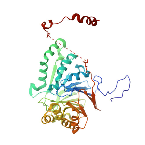
Structural Study of Metal Binding and Coordination in Ancient Metallo-beta-Lactamase PNGM-1 Variants.
Park, Y.S., Kim, T.Y., Park, H., Lee, J.H., Nguyen, D.Q., Hong, M.K., Lee, S.H., Kang, L.W.(2020) Int J Mol Sci 21
- PubMed: 32664695
- DOI: https://doi.org/10.3390/ijms21144926
- Primary Citation of Related Structures:
7BYQ, 7BZ1, 7BZ3, 7BZ4, 7BZI, 7WI1 - PubMed Abstract:
The increasing incidence of community- and hospital-acquired infections with multidrug-resistant (MDR) bacteria poses a critical threat to public health and the healthcare system. Although β-lactam antibiotics are effective against most bacterial infections, some bacteria are resistant to β-lactam antibiotics by producing β-lactamases. Among β-lactamases, metallo-β-lactamases (MBLs) are especially worrisome as only a few inhibitors have been developed against them. In MBLs, the metal ions play an important role as they coordinate a catalytic water molecule that hydrolyzes β-lactam rings. We determined the crystal structures of different variants of PNGM-1, an ancient MBL with additional tRNase Z activity. The variants were generated by site-directed mutagenesis targeting metal-coordinating residues. In PNGM-1, both zinc ions are coordinated by six coordination partners in an octahedral geometry, and the zinc-centered octahedrons share a common face. Structures of the PNGM-1 variants confirm that the substitution of a metal-coordinating residue causes the loss of metal binding and β-lactamase activity. Compared with PNGM-1, subclass B3 MBLs lack one metal-coordinating residue, leading to a shift in the metal-coordination geometry from an octahedral to tetrahedral geometry. Our results imply that a subtle change in the metal-binding site of MBLs can markedly change their metal-coordination geometry and catalytic activity.
Organizational Affiliation:
Department of Biological Sciences, Konkuk University, 120 Neungdong-ro, Gwangjin-gu, Seoul 05029, Korea.