7AO9

Structure of the core MTA1/HDAC1/MBD2 NURD deacetylase complex


Experimental Data Snapshot

  • Method: ELECTRON MICROSCOPY
  • Resolution: 6.10 Å
  • Aggregation State: PARTICLE 
  • Reconstruction Method: SINGLE PARTICLE 

Starting Models: experimental
View more details

wwPDB Validation   3D Report Full Report


This is version 1.2 of the entry. See complete history


Literature

The topology of chromatin-binding domains in the NuRD deacetylase complex.

Millard, C.J.Fairall, L.Ragan, T.J.Savva, C.G.Schwabe, J.W.R.

(2020) Nucleic Acids Res 48: 12972-12982

  • DOI: https://doi.org/10.1093/nar/gkaa1121
  • Primary Citation of Related Structures:  
    7AO8, 7AO9, 7AOA

  • PubMed Abstract: 

    Class I histone deacetylase complexes play essential roles in many nuclear processes. Whilst they contain a common catalytic subunit, they have diverse modes of action determined by associated factors in the distinct complexes. The deacetylase module from the NuRD complex contains three protein domains that control the recruitment of chromatin to the deacetylase enzyme, HDAC1/2. Using biochemical approaches and cryo-electron microscopy, we have determined how three chromatin-binding domains (MTA1-BAH, MBD2/3 and RBBP4/7) are assembled in relation to the core complex so as to facilitate interaction of the complex with the genome. We observe a striking arrangement of the BAH domains suggesting a potential mechanism for binding to di-nucleosomes. We also find that the WD40 domains from RBBP4 are linked to the core with surprising flexibility that is likely important for chromatin engagement. A single MBD2 protein binds asymmetrically to the dimerisation interface of the complex. This symmetry mismatch explains the stoichiometry of the complex. Finally, our structures suggest how the holo-NuRD might assemble on a di-nucleosome substrate.


  • Organizational Affiliation

    The Leicester Institute of Structural and Chemical Biology, Department of Molecular and Cell Biology, University of Leicester, Leicester LE1 7RH, UK.


Macromolecules
Find similar proteins by:  (by identity cutoff)  |  3D Structure
Entity ID: 1
MoleculeChains Sequence LengthOrganismDetailsImage
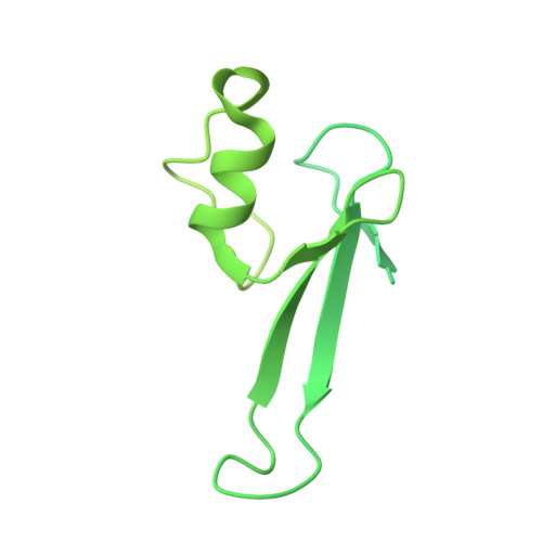
Methyl-CpG-binding domain protein 2A [auth C]411Homo sapiensMutation(s): 0 
Gene Names: MBD2
UniProt & NIH Common Fund Data Resources
Find proteins for Q9UBB5 (Homo sapiens)
Explore Q9UBB5 
Go to UniProtKB:  Q9UBB5
PHAROS:  Q9UBB5
GTEx:  ENSG00000134046 
Entity Groups  
Sequence Clusters30% Identity50% Identity70% Identity90% Identity95% Identity100% Identity
UniProt GroupQ9UBB5
Sequence Annotations
Expand
  • Reference Sequence
Find similar proteins by:  (by identity cutoff)  |  3D Structure
Entity ID: 2
MoleculeChains Sequence LengthOrganismDetailsImage
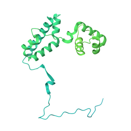
Metastasis-associated protein MTA1B [auth D],
D [auth A]
715Homo sapiensMutation(s): 0 
Gene Names: MTA1
UniProt & NIH Common Fund Data Resources
Find proteins for Q13330 (Homo sapiens)
Explore Q13330 
Go to UniProtKB:  Q13330
PHAROS:  Q13330
GTEx:  ENSG00000182979 
Entity Groups  
Sequence Clusters30% Identity50% Identity70% Identity90% Identity95% Identity100% Identity
UniProt GroupQ13330
Sequence Annotations
Expand
  • Reference Sequence
Find similar proteins by:  (by identity cutoff)  |  3D Structure
Entity ID: 3
MoleculeChains Sequence LengthOrganismDetailsImage
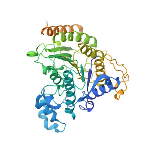
Histone deacetylase 1C [auth E],
E [auth B]
482Homo sapiensMutation(s): 0 
Gene Names: HDAC1RPD3L1
EC: 3.5.1.98 (PDB Primary Data), 3.5.1 (UniProt)
UniProt & NIH Common Fund Data Resources
Find proteins for Q13547 (Homo sapiens)
Explore Q13547 
Go to UniProtKB:  Q13547
PHAROS:  Q13547
GTEx:  ENSG00000116478 
Entity Groups  
Sequence Clusters30% Identity50% Identity70% Identity90% Identity95% Identity100% Identity
UniProt GroupQ13547
Sequence Annotations
Expand
  • Reference Sequence
Small Molecules
Ligands 3 Unique
IDChains Name / Formula / InChI Key2D Diagram3D Interactions
IHP
Query on IHP

Download Ideal Coordinates CCD File 
F [auth D],
J [auth A]
INOSITOL HEXAKISPHOSPHATE
C6 H18 O24 P6
IMQLKJBTEOYOSI-GPIVLXJGSA-N
ZN
Query on ZN

Download Ideal Coordinates CCD File 
G [auth E],
K [auth B]
ZINC ION
Zn
PTFCDOFLOPIGGS-UHFFFAOYSA-N
K
Query on K

Download Ideal Coordinates CCD File 
H [auth E],
I [auth E],
L [auth B],
M [auth B]
POTASSIUM ION
K
NPYPAHLBTDXSSS-UHFFFAOYSA-N
Experimental Data & Validation

Experimental Data

  • Method: ELECTRON MICROSCOPY
  • Resolution: 6.10 Å
  • Aggregation State: PARTICLE 
  • Reconstruction Method: SINGLE PARTICLE 
EM Software:
TaskSoftware PackageVersion
RECONSTRUCTIONRELION3.0

Structure Validation

View Full Validation Report


Entry History & Funding Information

Deposition Data


Funding OrganizationLocationGrant Number
Wellcome TrustUnited KingdomWT100237/Z/12/Z
Medical Research Council (MRC, United Kingdom)United KingdomMC_PC_17136
Wolfson FoundationUnited Kingdom--

Revision History  (Full details and data files)

  • Version 1.0: 2020-11-11
    Type: Initial release
  • Version 1.1: 2020-12-30
    Changes: Database references
  • Version 1.2: 2024-05-01
    Changes: Data collection, Database references, Refinement description