7ABN

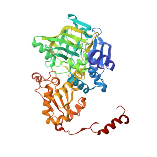
Structure of the reversible pyrrole-2-carboxylic acid decarboxylase PA0254/HudA


Experimental Data Snapshot

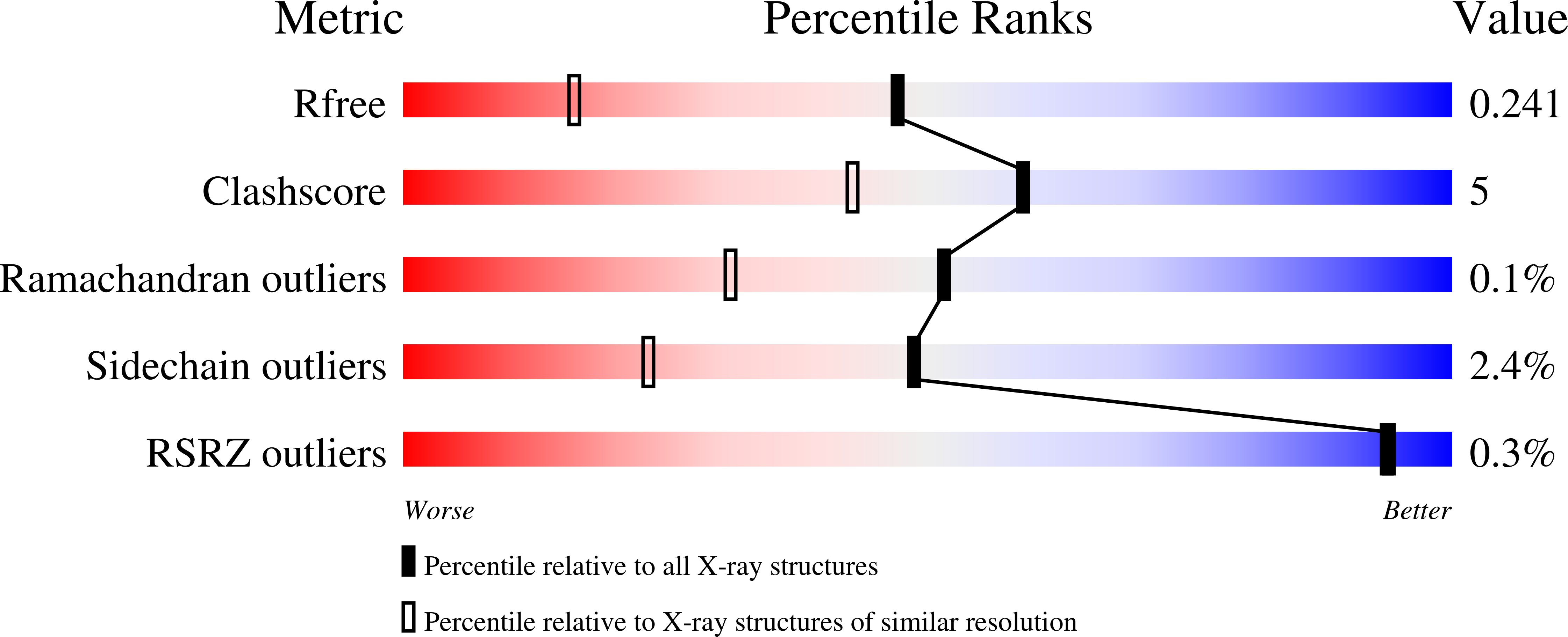
  • Method: X-RAY DIFFRACTION
  • Resolution: 1.65 Å
  • R-Value Free: 0.235 
  • R-Value Work: 0.201 
  • R-Value Observed: 0.203 

Starting Model: experimental
View more details

wwPDB Validation   3D Report Full Report


Ligand Structure Quality Assessment 


This is version 1.1 of the entry. See complete history


Literature

Structure and Mechanism of Pseudomonas aeruginosa PA0254/HudA, a prFMN-Dependent Pyrrole-2-carboxylic Acid Decarboxylase Linked to Virulence.

Payne, K.A.P.Marshall, S.A.Fisher, K.Rigby, S.E.J.Cliff, M.J.Spiess, R.Cannas, D.M.Larrosa, I.Hay, S.Leys, D.

(2021) ACS Catal 11: 2865-2878

  • DOI: https://doi.org/10.1021/acscatal.0c05042
  • Primary Citation of Related Structures:  
    7ABN, 7ABO

  • PubMed Abstract: 

    The UbiD family of reversible (de)carboxylases depends on the recently discovered prenylated-FMN (prFMN) cofactor for activity. The model enzyme ferulic acid decarboxylase (Fdc1) decarboxylates unsaturated aliphatic acids via a reversible 1,3-cycloaddition process. Protein engineering has extended the Fdc1 substrate range to include (hetero)aromatic acids, although catalytic rates remain poor. This raises the question how efficient decarboxylation of (hetero)aromatic acids is achieved by other UbiD family members. Here, we show that the Pseudomonas aeruginosa virulence attenuation factor PA0254 / HudA is a pyrrole-2-carboxylic acid decarboxylase. The crystal structure of the enzyme in the presence of the reversible inhibitor imidazole reveals a covalent prFMN - imidazole adduct is formed. Substrate screening reveals HudA and selected active site variants can accept a modest range of heteroaromatic compounds, including thiophene-2-carboxylic acid. Together with computational studies, our data suggests prFMN covalent catalysis occurs via electrophilic aromatic substitution and links HudA activity with the inhibitory effects of pyrrole-2-carboxylic acid on P. aeruginosa quorum sensing.


  • Organizational Affiliation

    Manchester Institute of Biotechnology, University of Manchester, 131 Princess Street, Manchester M1 7DN, United Kingdom.


Macromolecules
Find similar proteins by:  (by identity cutoff)  |  3D Structure
Entity ID: 1
MoleculeChains Sequence LengthOrganismDetailsImage
UbiD-like decarboxylaseA [auth D],
B [auth A],
C [auth H],
D [auth M]
496Pseudomonas aeruginosaMutation(s): 0 
Gene Names: ECC04_013425
EC: 4.1.1 (PDB Primary Data), 4.1.1.93 (UniProt)
UniProt
Find proteins for Q9I6N5 (Pseudomonas aeruginosa (strain ATCC 15692 / DSM 22644 / CIP 104116 / JCM 14847 / LMG 12228 / 1C / PRS 101 / PAO1))
Explore Q9I6N5 
Go to UniProtKB:  Q9I6N5
Entity Groups  
Sequence Clusters30% Identity50% Identity70% Identity90% Identity95% Identity100% Identity
UniProt GroupQ9I6N5
Sequence Annotations
Expand
  • Reference Sequence
Small Molecules
Ligands 4 Unique
IDChains Name / Formula / InChI Key2D Diagram3D Interactions
4LU (Subject of Investigation/LOI)
Query on 4LU

Download Ideal Coordinates CCD File 
G [auth D],
K [auth A],
O [auth H],
T [auth M]
1-deoxy-5-O-phosphono-1-(3,3,4,5-tetramethyl-9,11-dioxo-2,3,8,9,10,11-hexahydro-7H-quinolino[1,8-fg]pteridin-12-ium-7-y l)-D-ribitol
C22 H30 N4 O9 P
KOUJZPGFPGLHCZ-IYOUNJFTSA-O
IMD (Subject of Investigation/LOI)
Query on IMD

Download Ideal Coordinates CCD File 
H [auth D],
L [auth A],
P [auth H],
Q [auth M]
IMIDAZOLE
C3 H5 N2
RAXXELZNTBOGNW-UHFFFAOYSA-O
MN
Query on MN

Download Ideal Coordinates CCD File 
E [auth D],
I [auth A],
M [auth H],
R [auth M]
MANGANESE (II) ION
Mn
WAEMQWOKJMHJLA-UHFFFAOYSA-N
K
Query on K

Download Ideal Coordinates CCD File 
F [auth D],
J [auth A],
N [auth H],
S [auth M]
POTASSIUM ION
K
NPYPAHLBTDXSSS-UHFFFAOYSA-N
Experimental Data & Validation

Experimental Data

  • Method: X-RAY DIFFRACTION
  • Resolution: 1.65 Å
  • R-Value Free: 0.235 
  • R-Value Work: 0.201 
  • R-Value Observed: 0.203 
  • Space Group: P 1 21 1
Unit Cell:
Length ( Å )Angle ( ˚ )
a = 107.81α = 90
b = 55.48β = 99.92
c = 199.53γ = 90
Software Package:
Software NamePurpose
REFMACrefinement
xia2data reduction
xia2data scaling
PHASERphasing

Structure Validation

View Full Validation Report


Ligand Structure Quality Assessment 


Entry History & Funding Information

Deposition Data

  • Released Date: 2021-04-07 
  • Deposition Author(s): Leys, D.

Funding OrganizationLocationGrant Number
European Research Council (ERC)United KingdomADG_695013

Revision History  (Full details and data files)

  • Version 1.0: 2021-04-07
    Type: Initial release
  • Version 1.1: 2024-01-31
    Changes: Data collection, Database references, Refinement description