7XZ0

TRIM E3 ubiquitin ligase


Experimental Data Snapshot

  • Method: X-RAY DIFFRACTION
  • Resolution: 3.28 Å
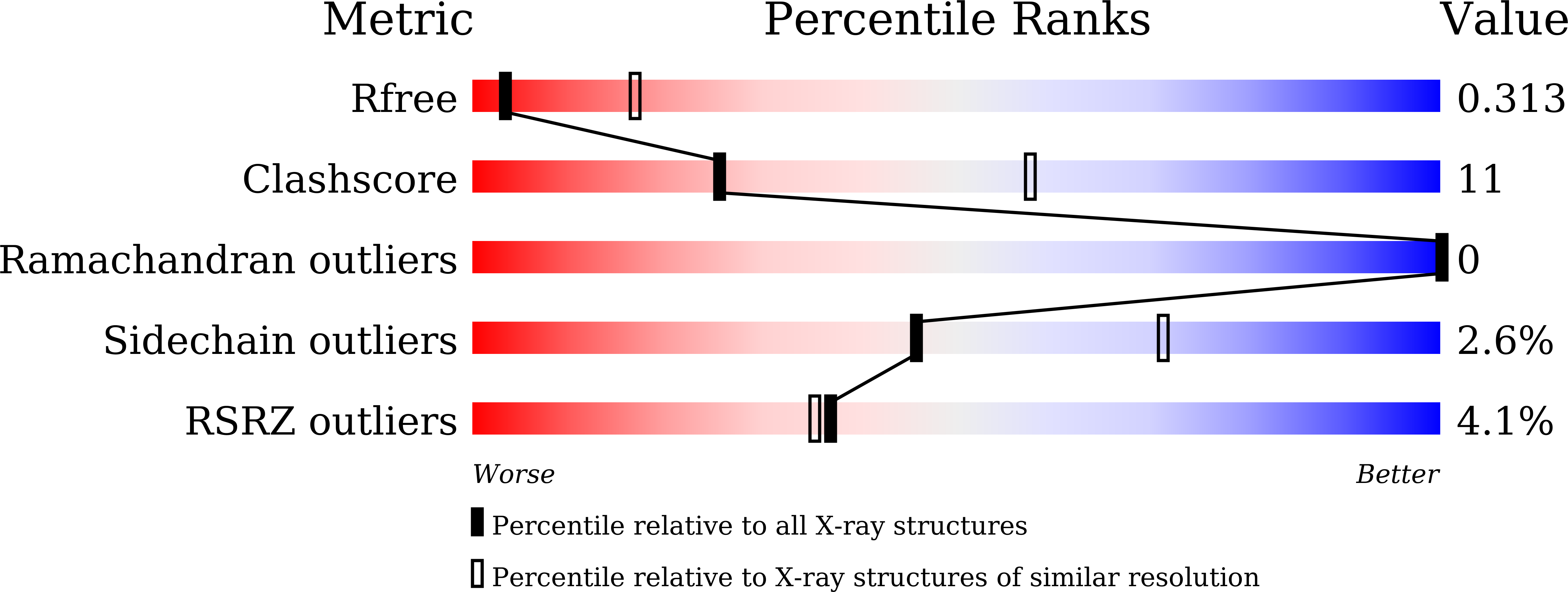
  • R-Value Free: 0.308 
  • R-Value Work: 0.254 
  • R-Value Observed: 0.259 

Starting Model: experimental
View more details

wwPDB Validation   3D Report Full Report


This is version 1.4 of the entry. See complete history


Literature

Structure and activation of the RING E3 ubiquitin ligase TRIM72 on the membrane.

Park, S.H.Han, J.Jeong, B.C.Song, J.H.Jang, S.H.Jeong, H.Kim, B.H.Ko, Y.G.Park, Z.Y.Lee, K.E.Hyun, J.Song, H.K.

(2023) Nat Struct Mol Biol 30: 1695-1706

  • DOI: https://doi.org/10.1038/s41594-023-01111-7
  • Primary Citation of Related Structures:  
    7XV2, 7XYY, 7XYZ, 7XZ0, 7XZ1, 7XZ2

  • PubMed Abstract: 

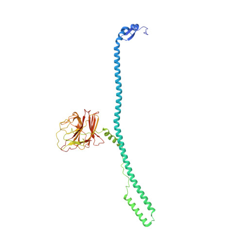
    Defects in plasma membrane repair can lead to muscle and heart diseases in humans. Tripartite motif-containing protein (TRIM)72 (mitsugumin 53; MG53) has been determined to rapidly nucleate vesicles at the site of membrane damage, but the underlying molecular mechanisms remain poorly understood. Here we present the structure of Mus musculus TRIM72, a complete model of a TRIM E3 ubiquitin ligase. We demonstrated that the interaction between TRIM72 and phosphatidylserine-enriched membranes is necessary for its oligomeric assembly and ubiquitination activity. Using cryogenic electron tomography and subtomogram averaging, we elucidated a higher-order model of TRIM72 assembly on the phospholipid bilayer. Combining structural and biochemical techniques, we developed a working molecular model of TRIM72, providing insights into the regulation of RING-type E3 ligases through the cooperation of multiple domains in higher-order assemblies. Our findings establish a fundamental basis for the study of TRIM E3 ligases and have therapeutic implications for diseases associated with membrane repair.


  • Organizational Affiliation

    Department of Life Sciences, Korea University, Seoul, South Korea.


Macromolecules
Find similar proteins by:  (by identity cutoff)  |  3D Structure
Entity ID: 1
MoleculeChains Sequence LengthOrganismDetailsImage
Tripartite motif-containing protein 72
A, B
394Mus musculusMutation(s): 4 
Gene Names: Trim72Mg53
EC: 2.3.2.27
UniProt
Find proteins for Q1XH17 (Mus musculus)
Explore Q1XH17 
Go to UniProtKB:  Q1XH17
Entity Groups  
Sequence Clusters30% Identity50% Identity70% Identity90% Identity95% Identity100% Identity
UniProt GroupQ1XH17
Sequence Annotations
Expand
  • Reference Sequence
Experimental Data & Validation

Experimental Data

  • Method: X-RAY DIFFRACTION
  • Resolution: 3.28 Å
  • R-Value Free: 0.308 
  • R-Value Work: 0.254 
  • R-Value Observed: 0.259 
  • Space Group: C 1 2 1
Unit Cell:
Length ( Å )Angle ( ˚ )
a = 239.728α = 90
b = 72.967β = 97.6
c = 83.764γ = 90
Software Package:
Software NamePurpose
PHENIXrefinement
HKL-2000data reduction
HKL-2000data scaling
PHASERphasing

Structure Validation

View Full Validation Report


Entry History & Funding Information

Deposition Data


Funding OrganizationLocationGrant Number
Other governmentKorea, Republic Of--

Revision History  (Full details and data files)

  • Version 1.0: 2023-07-12
    Type: Initial release
  • Version 1.1: 2023-10-04
    Changes: Data collection, Database references
  • Version 1.2: 2023-10-11
    Changes: Database references
  • Version 1.3: 2023-11-22
    Changes: Database references
  • Version 1.4: 2023-11-29
    Changes: Refinement description