7SO7

Novel structural insights for a pair of monoclonal antibodies recognizing non-overlapping epitopes of the glucosyltransferase domain of Clostridium difficile toxin B


Experimental Data Snapshot

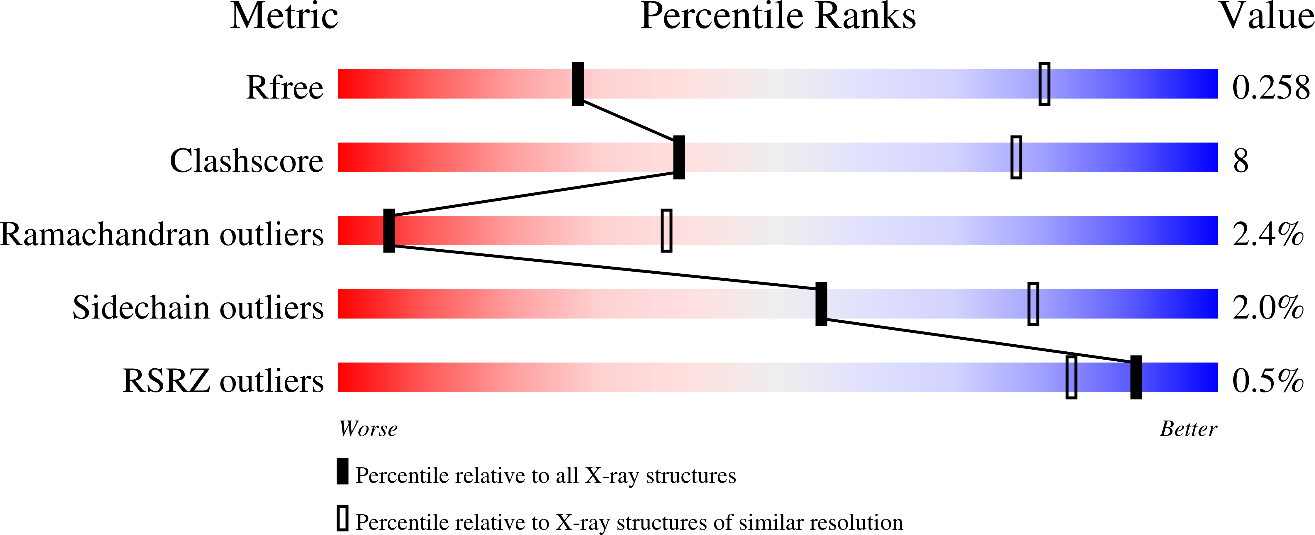
  • Method: X-RAY DIFFRACTION
  • Resolution: 3.59 Å
  • R-Value Free: 0.258 
  • R-Value Work: 0.209 
  • R-Value Observed: 0.211 

Starting Model: experimental
View more details

wwPDB Validation   3D Report Full Report


This is version 1.1 of the entry. See complete history


Literature

Novel structural insights for a pair of monoclonal antibodies recognizing non-overlapping epitopes of the glucosyltransferase domain of Clostridium difficile toxin B.

Liu, J.Kothe, M.Zhang, J.Oloo, E.Stegalkina, S.Mundle, S.T.Li, L.Zhang, J.Cole, L.E.Barone, L.Biemann, H.P.Kleanthous, H.Anosova, N.G.Anderson, S.F.

(2022) Curr Res Struct Biol 4: 96-105

  • DOI: https://doi.org/10.1016/j.crstbi.2022.03.003
  • Primary Citation of Related Structures:  
    7SO5, 7SO7

  • PubMed Abstract: 

    Clostridium difficile toxins are the primary causative agents for hospital-acquired diarrhea and pseudomembranous colitis. Numerous monoclonal antibodies (mAbs) targeting different domains of Clostridium difficile toxin have been reported. Here we report the crystal structures of two mAbs, B1 and B2, in complex with the glycosyltransferase domain (GTD) of the Clostridium difficile toxin B (TcdB). B2 bound to the N-terminal 4 helix bundle of the GTD, a conserved membrane localization domain (MLD) found in the large clostridial glycosylating toxin family implicated in targeting plasma membrane. B1 bound to a distinct epitope at the hinge region between the MLD and the catalytic subdomain of the GTD. Functional studies revealed the potency of these mAbs in vitro and in vivo to be synergistic when given in combination.


  • Organizational Affiliation

    Sanofi Integrated Drug Discovery, 153 2nd Ave, Waltham, 02451, USA.


Macromolecules
Find similar proteins by:  (by identity cutoff)  |  3D Structure
Entity ID: 1
MoleculeChains Sequence LengthOrganismDetailsImage
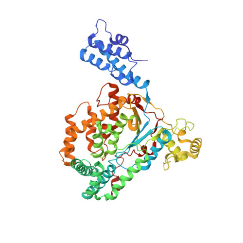
Toxin B
A, B
538Clostridioides difficileMutation(s): 0 
Gene Names: tcdBtoxB
EC: 3.4.22
UniProt
Find proteins for P18177 (Clostridioides difficile)
Explore P18177 
Go to UniProtKB:  P18177
Entity Groups  
Sequence Clusters30% Identity50% Identity70% Identity90% Identity95% Identity100% Identity
UniProt GroupP18177
Sequence Annotations
Expand
  • Reference Sequence
Find similar proteins by:  (by identity cutoff)  |  3D Structure
Entity ID: 2
MoleculeChains Sequence LengthOrganismDetailsImage
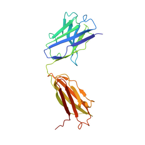
Fab B1 HCC [auth X],
E [auth H]
213Homo sapiensMutation(s): 0 
Entity Groups  
Sequence Clusters30% Identity50% Identity70% Identity90% Identity95% Identity100% Identity
Sequence Annotations
Expand
  • Reference Sequence
Find similar proteins by:  (by identity cutoff)  |  3D Structure
Entity ID: 3
MoleculeChains Sequence LengthOrganismDetailsImage
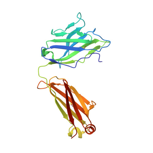
FAB B1 LCD [auth Y],
F [auth L]
218Homo sapiensMutation(s): 0 
Entity Groups  
Sequence Clusters30% Identity50% Identity70% Identity90% Identity95% Identity100% Identity
Sequence Annotations
Expand
  • Reference Sequence
Experimental Data & Validation

Experimental Data

  • Method: X-RAY DIFFRACTION
  • Resolution: 3.59 Å
  • R-Value Free: 0.258 
  • R-Value Work: 0.209 
  • R-Value Observed: 0.211 
  • Space Group: P 21 21 2
Unit Cell:
Length ( Å )Angle ( ˚ )
a = 210.54α = 90
b = 320.08β = 90
c = 65.61γ = 90
Software Package:
Software NamePurpose
PHENIXrefinement
Aimlessdata scaling
PDB_EXTRACTdata extraction
xia2data reduction
PHASERphasing

Structure Validation

View Full Validation Report


Entry History & Funding Information

Deposition Data

  • Released Date: 2022-05-11 
  • Deposition Author(s): Liu, J.

Funding OrganizationLocationGrant Number
Not funded--

Revision History  (Full details and data files)

  • Version 1.0: 2022-05-11
    Type: Initial release
  • Version 1.1: 2023-10-18
    Changes: Data collection, Refinement description