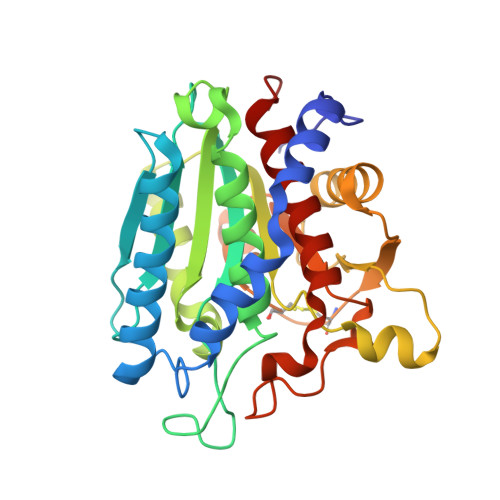
7OEZ

Leucine Aminopeptidase A mature enzyme in a complex with leucine


Experimental Data Snapshot

  • Method: X-RAY DIFFRACTION
  • Resolution: 2.48 Å
  • R-Value Free: 0.215 
  • R-Value Work: 0.157 
  • R-Value Observed: 0.160 

Starting Model: experimental
View more details

wwPDB Validation   3D Report Full Report


Ligand Structure Quality Assessment 


This is version 1.2 of the entry. See complete history


Literature

The role of propeptide-mediated autoinhibition and intermolecular chaperone in the maturation of cognate catalytic domain in leucine aminopeptidase.

Baltulionis, G.Blight, M.Robin, A.Charalampopoulos, D.Watson, K.A.

(2021) J Struct Biol 213: 107741-107741

  • DOI: https://doi.org/10.1016/j.jsb.2021.107741
  • Primary Citation of Related Structures:  
    6ZEP, 6ZEQ, 7OEZ

  • PubMed Abstract: 

    Leucyl aminopeptidase A from Aspergillus oryzae RIB40 (AO-LapA) is an exo-acting peptidase, widely utilised in food debittering applications. AO-LapA is secreted as a zymogen by the host and requires enzymatic cleavage of the autoinhibitory propeptide to reveal its full activity. Scarcity of structural data of zymogen aminopeptidases hampers a better understanding of the details of their molecular action of autoinhibition and how this might be utilised to improve the properties of such enzymes by recombinant methods for more effective bioprocessing. To address this gap in the literature, herein we report high-resolution crystal structures of recombinantly expressed AO-LapA precursor (AO-proLapA), mature LapA (AO-mLapA) and AO-mLapA complexed with reaction product l-leucine (AO-mLapA-Leu), all purified from Pichia pastoris culture supernatant. Our structures reveal a plausible molecular mechanism of LapA catalytic domain autoinhibition by propeptide and highlights the role of intramolecular chaperone (IMC). Our data suggest an absolute requirement for IMC in the maturation of cognate catalytic domain of AO-LapA. This observation is reinforced by our expression and refolding data of catalytic domain only (AO-refLapA) from Escherichia coli inclusion bodies, revealing a limited active conformation. Our work supports the notion that known synthetic aminopeptidase inhibitors and substrates mimic key polar contacts between propeptide and corresponding catalytic domain, demonstrated in our AO-proLapA zymogen crystal structure. Furthermore, understanding the atomic details of the autoinhibitory mechanism of cognate catalytic domains by native propeptides has wider reaching implications toward synthetic production of more effective inhibitors of bimetallic aminopeptidases and other dizinc enzymes that share an analogous reaction mechanism.


  • Organizational Affiliation

    School of Biological Science, Whiteknights Campus, University of Reading, RG6 6AS, UK; Department of Food and Nutritional Sciences, Whiteknights Campus, University of Reading, RG6 6AS, UK.


Macromolecules
Find similar proteins by:  (by identity cutoff)  |  3D Structure
Entity ID: 1
MoleculeChains Sequence LengthOrganismDetailsImage
Leucine aminopeptidase A301Aspergillus oryzae RIB40Mutation(s): 0 
Gene Names: lapAAO090011000052
EC: 3.4.11
UniProt
Find proteins for Q2U1F3 (Aspergillus oryzae (strain ATCC 42149 / RIB 40))
Explore Q2U1F3 
Go to UniProtKB:  Q2U1F3
Entity Groups  
Sequence Clusters30% Identity50% Identity70% Identity90% Identity95% Identity100% Identity
UniProt GroupQ2U1F3
Glycosylation
Glycosylation Sites: 1
Sequence Annotations
Expand
  • Reference Sequence
Oligosaccharides

Help

Entity ID: 2
MoleculeChains Length2D Diagram Glycosylation3D Interactions
2-acetamido-2-deoxy-beta-D-glucopyranose-(1-4)-2-acetamido-2-deoxy-beta-D-glucopyranose
B
2N-Glycosylation
Glycosylation Resources
GlyTouCan:  G42666HT
GlyCosmos:  G42666HT
GlyGen:  G42666HT
Small Molecules
Ligands 5 Unique
IDChains Name / Formula / InChI Key2D Diagram3D Interactions
LEU (Subject of Investigation/LOI)
Query on LEU

Download Ideal Coordinates CCD File 
P [auth A]LEUCINE
C6 H13 N O2
ROHFNLRQFUQHCH-YFKPBYRVSA-N
SO4
Query on SO4

Download Ideal Coordinates CCD File 
C [auth A],
D [auth A],
E [auth A],
F [auth A],
G [auth A]
SULFATE ION
O4 S
QAOWNCQODCNURD-UHFFFAOYSA-L
ZN
Query on ZN

Download Ideal Coordinates CCD File 
H [auth A],
I [auth A]
ZINC ION
Zn
PTFCDOFLOPIGGS-UHFFFAOYSA-N
EDO
Query on EDO

Download Ideal Coordinates CCD File 
K [auth A],
L [auth A],
M [auth A],
N [auth A],
O [auth A]
1,2-ETHANEDIOL
C2 H6 O2
LYCAIKOWRPUZTN-UHFFFAOYSA-N
CL
Query on CL

Download Ideal Coordinates CCD File 
J [auth A]CHLORIDE ION
Cl
VEXZGXHMUGYJMC-UHFFFAOYSA-M
Experimental Data & Validation

Experimental Data

  • Method: X-RAY DIFFRACTION
  • Resolution: 2.48 Å
  • R-Value Free: 0.215 
  • R-Value Work: 0.157 
  • R-Value Observed: 0.160 
  • Space Group: P 63 2 2
Unit Cell:
Length ( Å )Angle ( ˚ )
a = 154.1α = 90
b = 154.1β = 90
c = 87.83γ = 120
Software Package:
Software NamePurpose
PHENIXrefinement
PDB_EXTRACTdata extraction
APEXdata reduction
SCALAdata scaling
MOLREPphasing

Structure Validation

View Full Validation Report


Ligand Structure Quality Assessment 


Entry History & Funding Information

Deposition Data


Funding OrganizationLocationGrant Number
Biotechnology and Biological Sciences Research Council (BBSRC)United KingdomBB/K012053/1

Revision History  (Full details and data files)

  • Version 1.0: 2021-06-16
    Type: Initial release
  • Version 1.1: 2024-01-31
    Changes: Data collection, Database references, Refinement description
  • Version 1.2: 2024-11-13
    Changes: Structure summary