7K9M

Crystal structure of the complex of M. tuberculosis PheRS with cognate precursor tRNA and 5'-O-(N-phenylalanyl)sulfamoyl-adenosine


Experimental Data Snapshot

  • Method: X-RAY DIFFRACTION
  • Resolution: 2.50 Å
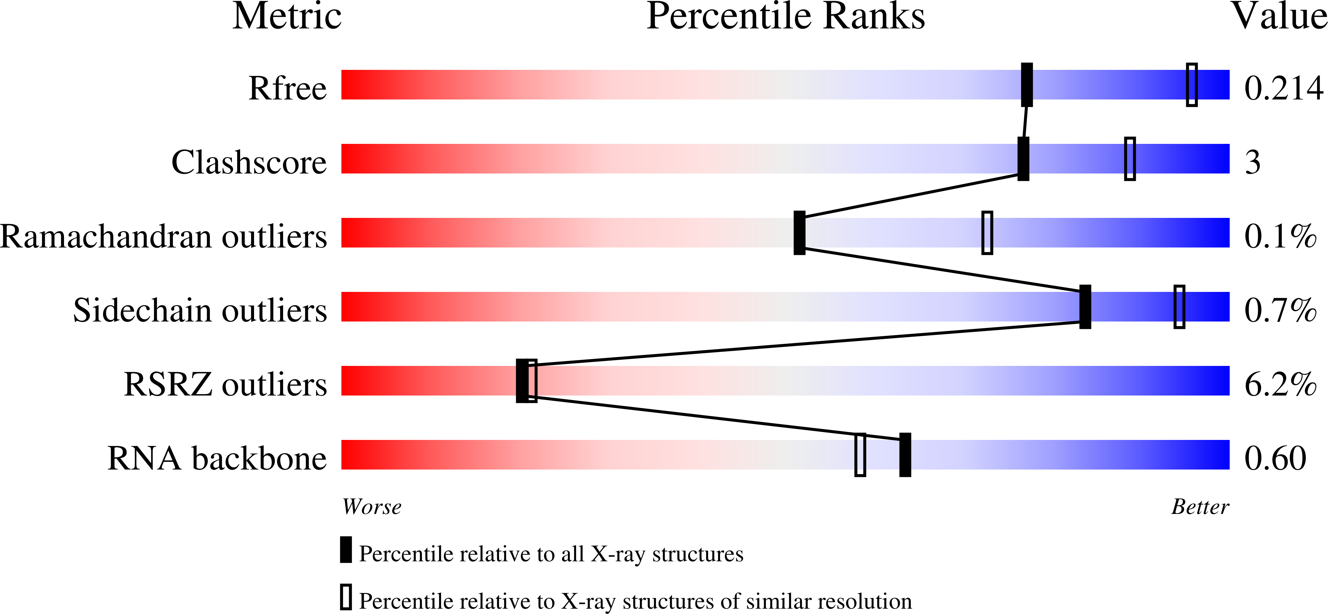
  • R-Value Free: 0.215 
  • R-Value Work: 0.179 
  • R-Value Observed: 0.181 

Starting Model: experimental
View more details

wwPDB Validation   3D Report Full Report


Ligand Structure Quality Assessment 


This is version 1.3 of the entry. See complete history


Literature

Mycobacterium tuberculosis Phe-tRNA synthetase: structural insights into tRNA recognition and aminoacylation.

Michalska, K.Jedrzejczak, R.Wower, J.Chang, C.Baragana, B.Gilbert, I.H.Forte, B.Joachimiak, A.

(2021) Nucleic Acids Res 49: 5351-5368

  • DOI: https://doi.org/10.1093/nar/gkab272
  • Primary Citation of Related Structures:  
    7K98, 7K9M, 7KA0, 7KAB

  • PubMed Abstract: 

    Tuberculosis, caused by Mycobacterium tuberculosis, responsible for ∼1.5 million fatalities in 2018, is the deadliest infectious disease. Global spread of multidrug resistant strains is a public health threat, requiring new treatments. Aminoacyl-tRNA synthetases are plausible candidates as potential drug targets, because they play an essential role in translating the DNA code into protein sequence by attaching a specific amino acid to their cognate tRNAs. We report structures of M. tuberculosis Phe-tRNA synthetase complexed with an unmodified tRNAPhe transcript and either L-Phe or a nonhydrolyzable phenylalanine adenylate analog. High-resolution models reveal details of two modes of tRNA interaction with the enzyme: an initial recognition via indirect readout of anticodon stem-loop and aminoacylation ready state involving interactions of the 3' end of tRNAPhe with the adenylate site. For the first time, we observe the protein gate controlling access to the active site and detailed geometry of the acyl donor and tRNA acceptor consistent with accepted mechanism. We biochemically validated the inhibitory potency of the adenylate analog and provide the most complete view of the Phe-tRNA synthetase/tRNAPhe system to date. The presented topography of amino adenylate-binding and editing sites at different stages of tRNA binding to the enzyme provide insights for the rational design of anti-tuberculosis drugs.


  • Organizational Affiliation

    Center for Structural Genomics of Infectious Diseases, Consortium for Advanced Science and Engineering, University of Chicago, Chicago, IL 60667, USA.


Macromolecules

Find similar proteins by:  (by identity cutoff)  |  3D Structure
Entity ID: 1
MoleculeChains Sequence LengthOrganismDetailsImage
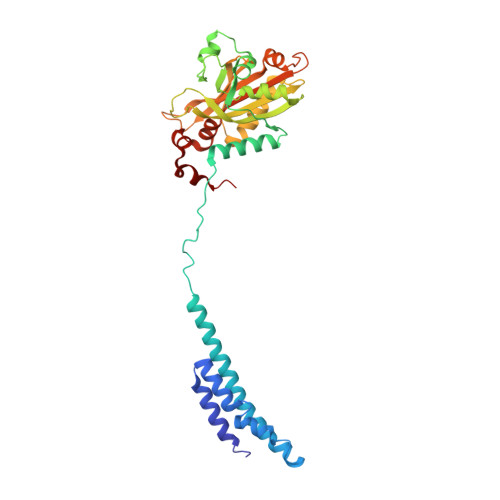
Phenylalanine--tRNA ligase alpha subunit341Mycobacterium tuberculosis H37RvMutation(s): 0 
Gene Names: pheSRv1649MTCY06H11.14
EC: 6.1.1.20
UniProt
Find proteins for P9WFU3 (Mycobacterium tuberculosis (strain ATCC 25618 / H37Rv))
Explore P9WFU3 
Go to UniProtKB:  P9WFU3
Entity Groups  
Sequence Clusters30% Identity50% Identity70% Identity90% Identity95% Identity100% Identity
UniProt GroupP9WFU3
Sequence Annotations
Expand
  • Reference Sequence
Find similar proteins by:  (by identity cutoff)  |  3D Structure
Entity ID: 2
MoleculeChains Sequence LengthOrganismDetailsImage
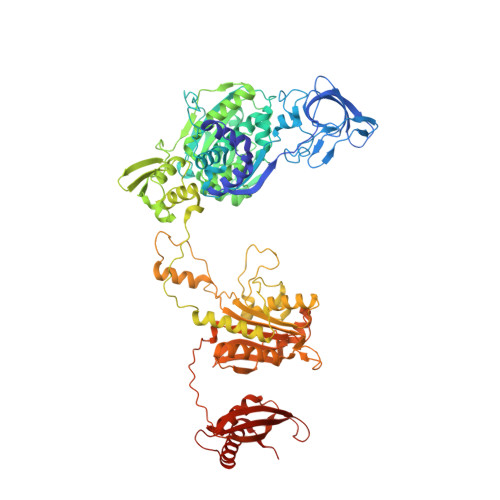
Phenylalanine--tRNA ligase beta subunit835Mycobacterium tuberculosis H37RvMutation(s): 0 
Gene Names: pheTRv1650MTCY06H11.15
EC: 6.1.1.20
UniProt
Find proteins for P9WFU1 (Mycobacterium tuberculosis (strain ATCC 25618 / H37Rv))
Explore P9WFU1 
Go to UniProtKB:  P9WFU1
Entity Groups  
Sequence Clusters30% Identity50% Identity70% Identity90% Identity95% Identity100% Identity
UniProt GroupP9WFU1
Sequence Annotations
Expand
  • Reference Sequence
Find similar nucleic acids by:  (by identity cutoff)  |  3D Structure
Entity ID: 3
MoleculeChains LengthOrganismImage
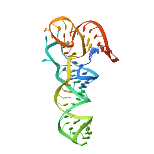
tRNA(Phe)77Mycobacterium tuberculosis H37Rv
Sequence Annotations
Expand
  • Reference Sequence
Small Molecules
Ligands 5 Unique
IDChains Name / Formula / InChI Key2D Diagram3D Interactions
W5Y (Subject of Investigation/LOI)
Query on W5Y

Download Ideal Coordinates CCD File 
D [auth A]5'-O-(L-phenylalanylsulfamoyl)adenosine
C19 H23 N7 O7 S
QGOJGCAOMCBQFJ-QTOWJTHWSA-N
P6G
Query on P6G

Download Ideal Coordinates CCD File 
L [auth B]HEXAETHYLENE GLYCOL
C12 H26 O7
IIRDTKBZINWQAW-UHFFFAOYSA-N
PGE
Query on PGE

Download Ideal Coordinates CCD File 
M [auth B]TRIETHYLENE GLYCOL
C6 H14 O4
ZIBGPFATKBEMQZ-UHFFFAOYSA-N
GOL
Query on GOL

Download Ideal Coordinates CCD File 
G [auth A]
H [auth A]
I [auth A]
J [auth A]
N [auth B]
G [auth A],
H [auth A],
I [auth A],
J [auth A],
N [auth B],
O [auth B],
P [auth B]
GLYCEROL
C3 H8 O3
PEDCQBHIVMGVHV-UHFFFAOYSA-N
MG
Query on MG

Download Ideal Coordinates CCD File 
E [auth A],
F [auth A],
K [auth A]
MAGNESIUM ION
Mg
JLVVSXFLKOJNIY-UHFFFAOYSA-N
Experimental Data & Validation

Experimental Data

Unit Cell:
Length ( Å )Angle ( ˚ )
a = 128.432α = 90
b = 110.753β = 103.59
c = 147.897γ = 90
Software Package:
Software NamePurpose
PHENIXrefinement
SCALEPACKdata scaling
PDB_EXTRACTdata extraction
HKL-3000data reduction
PHENIXphasing
SBC-Collectdata collection

Structure Validation

View Full Validation Report


Ligand Structure Quality Assessment 


Entry History & Funding Information

Deposition Data


Funding OrganizationLocationGrant Number
National Institutes of Health/National Institute Of Allergy and Infectious Diseases (NIH/NIAID)United StatesHHSN272201700060C

Revision History  (Full details and data files)

  • Version 1.0: 2021-05-12
    Type: Initial release
  • Version 1.1: 2021-06-02
    Changes: Database references
  • Version 1.2: 2021-06-16
    Changes: Database references
  • Version 1.3: 2023-10-18
    Changes: Data collection, Database references, Refinement description