7CZ4

Structure of SARS-CoV-2 macro domain in complex with ADP-ribose


Experimental Data Snapshot

  • Method: X-RAY DIFFRACTION
  • Resolution: 2.64 Å
  • R-Value Free: 0.215 
  • R-Value Work: 0.171 
  • R-Value Observed: 0.175 

Starting Model: experimental
View more details

wwPDB Validation   3D Report Full Report


Ligand Structure Quality Assessment 


This is version 1.3 of the entry. See complete history


Literature

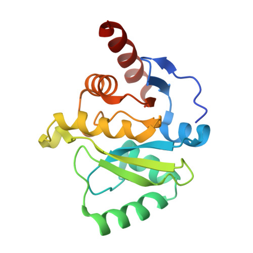
Structural, Biophysical, and Biochemical Elucidation of the SARS-CoV-2 Nonstructural Protein 3 Macro Domain.

Lin, M.H.Chang, S.C.Chiu, Y.C.Jiang, B.C.Wu, T.H.Hsu, C.H.

(2020) ACS Infect Dis 6: 2970-2978

  • DOI: https://doi.org/10.1021/acsinfecdis.0c00441
  • Primary Citation of Related Structures:  
    7C33, 7CZ4

  • PubMed Abstract: 

    The pandemic outbreak of a novel coronavirus, severe acute respiratory syndrome coronavirus 2 (SARS-CoV-2), has threatened the global public health and economy since late December 2019. SARS-CoV-2 encodes the conserved macro domain within nonstructural protein 3, which may reverse cellular ADP-ribosylation and potentially cut the signal of a viral infection in the cell. Herein, we report that the SARS-CoV-2 macro domain was examined as a poly-ADP-ribose (ADPR) binding module and possessed mono-ADPR cleavage enzyme activity. After confirming the ADPR binding ability via a biophysical approach, the X-ray crystal structure of the SARS-CoV-2 macro domain was determined and structurally compared with those of other viruses. This study provides structural, biophysical, and biochemical bases to further evaluate the role of the SARS-CoV-2 macro domain in the host response via ADP-ribose binding but also as a potential target for drug design against COVID-19.


  • Organizational Affiliation

    Genome and Systems Biology Degree Program, National Taiwan University and Academia Sinica, Taipei 10617, Taiwan.


Macromolecules
Find similar proteins by:  (by identity cutoff)  |  3D Structure
Entity ID: 1
MoleculeChains Sequence LengthOrganismDetailsImage
Non-structural protein 3
A, B
175Severe acute respiratory syndrome coronavirus 2Mutation(s): 0 
EC: 3.4.19.12
UniProt
Find proteins for P0DTD1 (Severe acute respiratory syndrome coronavirus 2)
Explore P0DTD1 
Go to UniProtKB:  P0DTD1
Entity Groups  
Sequence Clusters30% Identity50% Identity70% Identity90% Identity95% Identity100% Identity
UniProt GroupP0DTD1
Sequence Annotations
Expand
  • Reference Sequence
Experimental Data & Validation

Experimental Data

  • Method: X-RAY DIFFRACTION
  • Resolution: 2.64 Å
  • R-Value Free: 0.215 
  • R-Value Work: 0.171 
  • R-Value Observed: 0.175 
  • Space Group: P 41 21 2
Unit Cell:
Length ( Å )Angle ( ˚ )
a = 84.736α = 90
b = 84.736β = 90
c = 143.242γ = 90
Software Package:
Software NamePurpose
PHENIXrefinement
HKL-2000data reduction
HKL-2000data scaling
PHASERphasing

Structure Validation

View Full Validation Report


Ligand Structure Quality Assessment 


Entry History & Funding Information

Deposition Data


Funding OrganizationLocationGrant Number
Ministry of Science and Technology (MoST, Taiwan)Taiwan108-2628-B-002-013
Ministry of Science and Technology (MoST, Taiwan)Taiwan108-2113-M-002-011

Revision History  (Full details and data files)

  • Version 1.0: 2020-11-11
    Type: Initial release
  • Version 1.1: 2020-12-02
    Changes: Database references
  • Version 1.2: 2021-03-10
    Changes: Structure summary
  • Version 1.3: 2023-11-29
    Changes: Data collection, Database references, Refinement description