6Z6D

Crystal structure of the HK97 bacteriophage large terminase


Experimental Data Snapshot

  • Method: X-RAY DIFFRACTION
  • Resolution: 2.20 Å
  • R-Value Free: 0.254 
  • R-Value Work: 0.205 
  • R-Value Observed: 0.207 

wwPDB Validation   3D Report Full Report


This is version 1.2 of the entry. See complete history


Literature

Structural basis of DNA packaging by a ring-type ATPase from an archetypal viral system.

Fung, H.K.H.Grimes, S.Huet, A.Duda, R.L.Chechik, M.Gault, J.Robinson, C.V.Hendrix, R.W.Jardine, P.J.Conway, J.F.Baumann, C.G.Antson, A.A.

(2022) Nucleic Acids Res 50: 8719-8732

  • DOI: https://doi.org/10.1093/nar/gkac647
  • Primary Citation of Related Structures:  
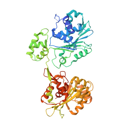
    6Z6D, 6Z6E

  • PubMed Abstract: 

    Many essential cellular processes rely on substrate rotation or translocation by a multi-subunit, ring-type NTPase. A large number of double-stranded DNA viruses, including tailed bacteriophages and herpes viruses, use a homomeric ring ATPase to processively translocate viral genomic DNA into procapsids during assembly. Our current understanding of viral DNA packaging comes from three archetypal bacteriophage systems: cos, pac and phi29. Detailed mechanistic understanding exists for pac and phi29, but not for cos. Here, we reconstituted in vitro a cos packaging system based on bacteriophage HK97 and provided a detailed biochemical and structural description. We used a photobleaching-based, single-molecule assay to determine the stoichiometry of the DNA-translocating ATPase large terminase. Crystal structures of the large terminase and DNA-recruiting small terminase, a first for a biochemically defined cos system, reveal mechanistic similarities between cos and pac systems. At the same time, mutational and biochemical analyses indicate a new regulatory mechanism for ATPase multimerization and coordination in the HK97 system. This work therefore establishes a framework for studying the evolutionary relationships between ATP-dependent DNA translocation machineries in double-stranded DNA viruses.


  • Organizational Affiliation

    Department of Biology, University of York, York, YO10 5DD, UK.


Macromolecules
Find similar proteins by:  (by identity cutoff)  |  3D Structure
Entity ID: 1
MoleculeChains Sequence LengthOrganismDetailsImage
Terminase large subunit514Byrnievirus HK97Mutation(s): 0 
UniProt
Find proteins for Q9MCT1 (Enterobacteria phage HK97)
Explore Q9MCT1 
Go to UniProtKB:  Q9MCT1
Entity Groups  
Sequence Clusters30% Identity50% Identity70% Identity90% Identity95% Identity100% Identity
UniProt GroupQ9MCT1
Sequence Annotations
Expand
  • Reference Sequence
Small Molecules
Ligands 1 Unique
IDChains Name / Formula / InChI Key2D Diagram3D Interactions
BR
Query on BR

Download Ideal Coordinates CCD File 
B [auth A]
C [auth A]
D [auth A]
E [auth A]
F [auth A]
B [auth A],
C [auth A],
D [auth A],
E [auth A],
F [auth A],
G [auth A],
H [auth A],
I [auth A],
J [auth A],
K [auth A],
L [auth A],
M [auth A],
N [auth A],
O [auth A],
P [auth A],
Q [auth A],
R [auth A]
BROMIDE ION
Br
CPELXLSAUQHCOX-UHFFFAOYSA-M
Experimental Data & Validation

Experimental Data

  • Method: X-RAY DIFFRACTION
  • Resolution: 2.20 Å
  • R-Value Free: 0.254 
  • R-Value Work: 0.205 
  • R-Value Observed: 0.207 
  • Space Group: C 1 2 1
Unit Cell:
Length ( Å )Angle ( ˚ )
a = 211.787α = 90
b = 39.146β = 103.39
c = 65.45γ = 90
Software Package:
Software NamePurpose
REFMACrefinement
PDB_EXTRACTdata extraction
XDSdata reduction
XDSdata scaling
SHELXDphasing

Structure Validation

View Full Validation Report


Entry History & Funding Information

Deposition Data


Funding OrganizationLocationGrant Number
Wellcome TrustUnited Kingdom098230
Wellcome TrustUnited Kingdom095024MA

Revision History  (Full details and data files)

  • Version 1.0: 2021-06-09
    Type: Initial release
  • Version 1.1: 2022-12-21
    Changes: Database references
  • Version 1.2: 2024-06-19
    Changes: Data collection