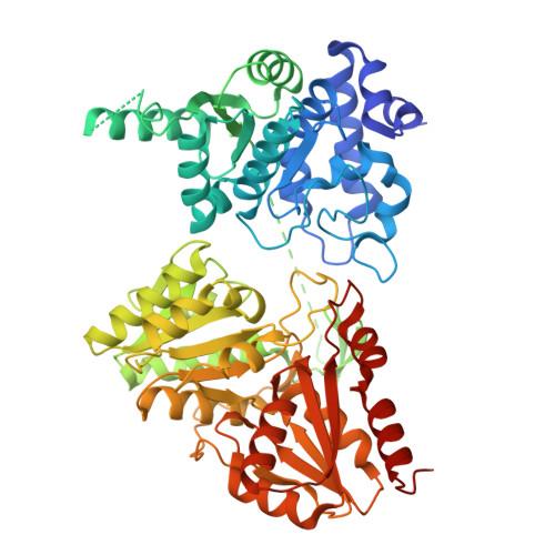
6XXG

Structure of truncated 1-deoxy-D-xylulose 5-phosphate synthase (DXS) from Deinococcus radiodurans


Experimental Data Snapshot

  • Method: X-RAY DIFFRACTION
  • Resolution: 2.10 Å
  • R-Value Free: 0.236 
  • R-Value Work: 0.191 

Starting Model: experimental
View more details

wwPDB Validation   3D Report Full Report


Ligand Structure Quality Assessment 


This is version 1.1 of the entry. See complete history


Literature

Identification of a 1-deoxy-D-xylulose-5-phosphate synthase (DXS) mutant with improved crystallographic properties.

Gierse, R.M.Reddem, E.R.Alhayek, A.Baitinger, D.Hamid, Z.Jakobi, H.Laber, B.Lange, G.Hirsch, A.K.H.Groves, M.R.

(2021) Biochem Biophys Res Commun 539: 42-47

  • DOI: https://doi.org/10.1016/j.bbrc.2020.12.069
  • Primary Citation of Related Structures:  
    6XXG

  • PubMed Abstract: 

    In this report, we describe a truncated Deinococcus radiodurans 1-deoxy-D-xylulose-5-phosphate synthase (DXS) protein that retains enzymatic activity, while slowing protein degradation and showing improved crystallization properties. With modern drug-design approaches relying heavily on the elucidation of atomic interactions of potential new drugs with their targets, the need for co-crystal structures with the compounds of interest is high. DXS itself is a promising drug target, as it catalyzes the first reaction in the 2-C-methyl-D-erythritol 4-phosphate (MEP)-pathway for the biosynthesis of the universal precursors of terpenes, which are essential secondary metabolites. In contrast to many bacteria and pathogens, which employ the MEP pathway, mammals use the distinct mevalonate-pathway for the biosynthesis of these precursors, which makes all enzymes of the MEP-pathway potential new targets for the development of anti-infectives. However, crystallization of DXS has proven to be challenging: while the first X-ray structures from Escherichia coli and D. radiodurans were solved in 2004, since then only two additions have been made in 2019 that were obtained under anoxic conditions. The presented site of truncation can potentially also be transferred to other homologues, opening up the possibility for the determination of crystal structures from pathogenic species, which until now could not be crystallized. This manuscript also provides a further example that truncation of a variable region of a protein can lead to improved structural data.


  • Organizational Affiliation

    Department for Drug Design and Optimization, Helmholtz Institute for Pharmaceutical Research (HIPS) - Helmholtz Centre for Infection Research (HZI), Campus Building E 8.1, 66123, Saarbrücken, Germany; Department of Pharmacy, Saarland University, Campus Building E8.1, 66123, Saarbrücken, Germany; Stratingh Institute for Chemistry, University of Groningen, Nijenborgh 7, 9747, AG Groningen, Netherlands.


Macromolecules
Find similar proteins by:  (by identity cutoff)  |  3D Structure
Entity ID: 1
MoleculeChains Sequence LengthOrganismDetailsImage
1-deoxy-D-xylulose-5-phosphate synthase,1-deoxy-D-xylulose-5-phosphate synthaseA [auth AAA],
B [auth BBB]
594Deinococcus radiodurans R1 = ATCC 13939 = DSM 20539Mutation(s): 0 
Gene Names: dxsDR_1475
EC: 2.2.1.7
UniProt
Find proteins for Q9RUB5 (Deinococcus radiodurans (strain ATCC 13939 / DSM 20539 / JCM 16871 / CCUG 27074 / LMG 4051 / NBRC 15346 / NCIMB 9279 / VKM B-1422 / R1))
Explore Q9RUB5 
Go to UniProtKB:  Q9RUB5
Entity Groups  
Sequence Clusters30% Identity50% Identity70% Identity90% Identity95% Identity100% Identity
UniProt GroupQ9RUB5
Sequence Annotations
Expand
  • Reference Sequence
Small Molecules
Ligands 3 Unique
IDChains Name / Formula / InChI Key2D Diagram3D Interactions
TPP (Subject of Investigation/LOI)
Query on TPP

Download Ideal Coordinates CCD File 
E [auth AAA],
L [auth BBB]
THIAMINE DIPHOSPHATE
C12 H19 N4 O7 P2 S
AYEKOFBPNLCAJY-UHFFFAOYSA-O
CA
Query on CA

Download Ideal Coordinates CCD File 
D [auth AAA]
F [auth AAA]
I [auth BBB]
M [auth BBB]
N [auth BBB]
D [auth AAA],
F [auth AAA],
I [auth BBB],
M [auth BBB],
N [auth BBB],
O [auth BBB]
CALCIUM ION
Ca
BHPQYMZQTOCNFJ-UHFFFAOYSA-N
MG (Subject of Investigation/LOI)
Query on MG

Download Ideal Coordinates CCD File 
C [auth AAA],
G [auth AAA],
H [auth BBB],
J [auth BBB],
K [auth BBB]
MAGNESIUM ION
Mg
JLVVSXFLKOJNIY-UHFFFAOYSA-N
Experimental Data & Validation

Experimental Data

  • Method: X-RAY DIFFRACTION
  • Resolution: 2.10 Å
  • R-Value Free: 0.236 
  • R-Value Work: 0.191 
  • Space Group: P 21 21 21
Unit Cell:
Length ( Å )Angle ( ˚ )
a = 72.321α = 90
b = 87.367β = 90
c = 181.865γ = 90
Software Package:
Software NamePurpose
REFMACrefinement
XDSdata reduction
XDSdata scaling
MOLREPphasing
Cootmodel building

Structure Validation

View Full Validation Report


Ligand Structure Quality Assessment 


Entry History & Funding Information

Deposition Data


Funding OrganizationLocationGrant Number
Netherlands Organisation for Scientific ResearchNetherlands731.015.414

Revision History  (Full details and data files)

  • Version 1.0: 2021-01-20
    Type: Initial release
  • Version 1.1: 2024-01-24
    Changes: Data collection, Database references, Derived calculations, Refinement description