


Generalized enzymatic mechanism of catalysis by tetrameric L-asparaginases from mesophilic bacteria.
Strzelczyk, P., Zhang, D., Dyba, M., Wlodawer, A., Lubkowski, J.(2020) Sci Rep 10: 17516
Experimental Data Snapshot
Starting Model: experimental
View more details
(2020) Sci Rep 10: 17516
Entity ID: 1 | |||||
|---|---|---|---|---|---|
| Molecule | Chains | Sequence Length | Organism | Details | Image |
| Glutaminase-asparaginase | 337 | Pseudomonas putida KT2440 | Mutation(s): 0 Gene Names: ansB, PP_2453 EC: 3.5.1.38 | 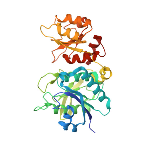 | |
UniProt | |||||
Find proteins for Q88K39 (Pseudomonas putida (strain ATCC 47054 / DSM 6125 / CFBP 8728 / NCIMB 11950 / KT2440)) Explore Q88K39 Go to UniProtKB: Q88K39 | |||||
Entity Groups | |||||
| Sequence Clusters | 30% Identity50% Identity70% Identity90% Identity95% Identity100% Identity | ||||
| UniProt Group | Q88K39 | ||||
Sequence AnnotationsExpand | |||||
| |||||
| Ligands 3 Unique | |||||
|---|---|---|---|---|---|
| ID | Chains | Name / Formula / InChI Key | 2D Diagram | 3D Interactions | |
| GLU (Subject of Investigation/LOI) Query on GLU | E [auth A], H [auth B], K [auth C], N [auth D] | GLUTAMIC ACID C5 H9 N O4 WHUUTDBJXJRKMK-VKHMYHEASA-N |  | ||
| GOL Query on GOL | I [auth B] | GLYCEROL C3 H8 O3 PEDCQBHIVMGVHV-UHFFFAOYSA-N |  | ||
| EDO Query on EDO | F [auth A] G [auth A] J [auth B] L [auth C] M [auth C] | 1,2-ETHANEDIOL C2 H6 O2 LYCAIKOWRPUZTN-UHFFFAOYSA-N |  | ||
| Length ( Å ) | Angle ( ˚ ) |
|---|---|
| a = 81.542 | α = 90 |
| b = 130.791 | β = 117.82 |
| c = 81.548 | γ = 90 |
| Software Name | Purpose |
|---|---|
| HKL-3000 | data scaling |
| REFMAC | refinement |
| PDB_EXTRACT | data extraction |
| HKL-3000 | data reduction |
| PHASER | phasing |
| Funding Organization | Location | Grant Number |
|---|---|---|
| National Institutes of Health/National Cancer Institute (NIH/NCI) | United States | -- |
RCSB PDB (citation) is hosted by
RCSB PDB is a member of the

