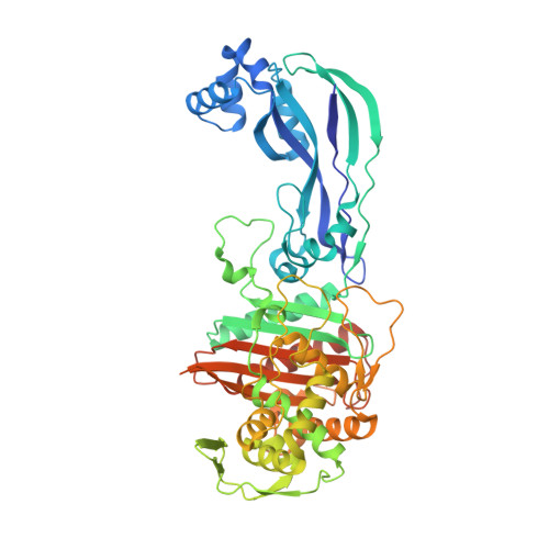
6VOT

Crystal structure of Pseudomonas aerugonisa PBP3 complexed to gamma-lactam YU253434


Experimental Data Snapshot

  • Method: X-RAY DIFFRACTION
  • Resolution: 2.40 Å
  • R-Value Free: 0.236 
  • R-Value Work: 0.182 
  • R-Value Observed: 0.185 

Starting Model: experimental
View more details

wwPDB Validation   3D Report Full Report

Currently 6VOT does not have a validation slider image.


This is version 1.3 of the entry. See complete history


Literature

A gamma-Lactam Siderophore Antibiotic Effective against Multidrug-Resistant Gram-Negative Bacilli.

Goldberg, J.A.Nguyen, H.Kumar, V.Spencer, E.J.Hoyer, D.Marshall, E.K.Cmolik, A.O'Shea, M.Marshall, S.H.Hujer, A.M.Hujer, K.M.Rudin, S.D.Domitrovic, T.N.Bethel, C.R.Papp-Wallace, K.M.Logan, L.K.Perez, F.Jacobs, M.R.van Duin, D.Kreiswirth, B.M.Bonomo, R.A.Plummer, M.S.van den Akker, F.

(2020) J Med Chem 63: 5990-6002

  • DOI: https://doi.org/10.1021/acs.jmedchem.0c00255
  • Primary Citation of Related Structures:  
    6VOT

  • PubMed Abstract: 

    Treatment of multidrug-resistant Gram-negative bacterial pathogens represents a critical clinical need. Here, we report a novel γ-lactam pyrazolidinone that targets penicillin-binding proteins (PBPs) and incorporates a siderophore moiety to facilitate uptake into the periplasm. The MIC values of γ-lactam YU253434, 1 , are reported along with the finding that 1 is resistant to hydrolysis by all four classes of β-lactamases. The druglike characteristics and mouse PK data are described along with the X-ray crystal structure of 1 binding to its target PBP3.


  • Organizational Affiliation

    Research Service, Louis Stokes Cleveland Department of Veterans Affairs Medical Center, Cleveland, Ohio 44106, United States.


Macromolecules
Find similar proteins by:  (by identity cutoff)  |  3D Structure
Entity ID: 1
MoleculeChains Sequence LengthOrganismDetailsImage
Peptidoglycan D,D-transpeptidase FtsI538Pseudomonas aeruginosaMutation(s): 0 
Gene Names: 
EC: 3.4.16.4
UniProt
Find proteins for G3XD46 (Pseudomonas aeruginosa (strain ATCC 15692 / DSM 22644 / CIP 104116 / JCM 14847 / LMG 12228 / 1C / PRS 101 / PAO1))
Explore G3XD46 
Go to UniProtKB:  G3XD46
Entity Groups  
Sequence Clusters30% Identity50% Identity70% Identity90% Identity95% Identity100% Identity
UniProt GroupG3XD46
Sequence Annotations
Expand
  • Reference Sequence
Small Molecules
Ligands 1 Unique
IDChains Name / Formula / InChI Key2D Diagram3D Interactions
R7G (Subject of Investigation/LOI)
Query on R7G

Download Ideal Coordinates CCD File 
B [auth A]1-[(2S)-2-{[(2Z)-2-(2-amino-1,3-thiazol-4-yl)-2-{[(2-carboxypropan-2-yl)oxy]imino}acetyl]amino}-3-oxopropyl]-4-{[2-(5,6 -dihydroxy-1,3-dioxo-1,3-dihydro-2H-isoindol-2-yl)ethyl]carbamoyl}-2,5-dihydro-1H-pyrazole-3-carboxylic acid
C27 H28 N8 O12 S
AUDGPVZSFLGMAE-ZJIUGIBFSA-N
Experimental Data & Validation

Experimental Data

  • Method: X-RAY DIFFRACTION
  • Resolution: 2.40 Å
  • R-Value Free: 0.236 
  • R-Value Work: 0.182 
  • R-Value Observed: 0.185 
  • Space Group: P 21 21 21
Unit Cell:
Length ( Å )Angle ( ˚ )
a = 68.107α = 90
b = 83.813β = 90
c = 89.74γ = 90
Software Package:
Software NamePurpose
DENZOdata reduction
HKL-2000data scaling
REFMACrefinement
PDB_EXTRACTdata extraction
PHASERphasing

Structure Validation

View Full Validation Report

Currently 6VOT does not have a validation slider image.


Entry History 

Deposition Data

Revision History  (Full details and data files)

  • Version 1.0: 2020-05-13
    Type: Initial release
  • Version 1.1: 2020-07-01
    Changes: Database references, Derived calculations, Structure summary
  • Version 1.2: 2023-10-11
    Changes: Data collection, Database references, Refinement description
  • Version 1.3: 2024-10-23
    Changes: Structure summary