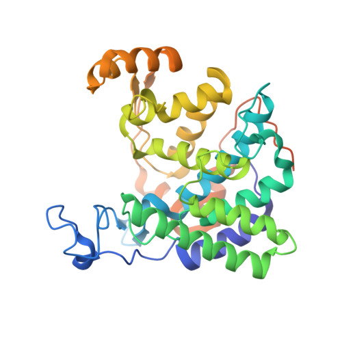
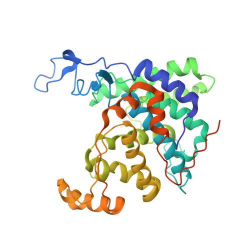
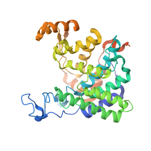
6RYB

Structure of deubiquitinase for PR-ubiquitination 1 -Dup1


Experimental Data Snapshot

  • Method: X-RAY DIFFRACTION
  • Resolution: 2.31 Å
  • R-Value Free: 0.247 
  • R-Value Work: 0.217 
  • R-Value Observed: 0.218 

Starting Model: experimental
View more details

wwPDB Validation   3D Report Full Report


This is version 1.3 of the entry. See complete history


Literature

Regulation of Phosphoribosyl-Linked Serine Ubiquitination by Deubiquitinases DupA and DupB.

Shin, D.Mukherjee, R.Liu, Y.Gonzalez, A.Bonn, F.Liu, Y.Rogov, V.V.Heinz, M.Stolz, A.Hummer, G.Dotsch, V.Luo, Z.Q.Bhogaraju, S.Dikic, I.

(2020) Mol Cell 77: 164-179.e6

  • DOI: https://doi.org/10.1016/j.molcel.2019.10.019
  • Primary Citation of Related Structures:  
    6RYA, 6RYB

  • PubMed Abstract: 

    The family of bacterial SidE enzymes catalyzes non-canonical phosphoribosyl-linked (PR) serine ubiquitination and promotes infectivity of Legionella pneumophila. Here, we describe identification of two bacterial effectors that reverse PR ubiquitination and are thus named deubiquitinases for PR ubiquitination (DUPs; DupA and DupB). Structural analyses revealed that DupA and SidE ubiquitin ligases harbor a highly homologous catalytic phosphodiesterase (PDE) domain. However, unlike SidE ubiquitin ligases, DupA displays increased affinity to PR-ubiquitinated substrates, which allows DupA to cleave PR ubiquitin from substrates. Interfering with DupA-ubiquitin binding switches its activity toward SidE-type ligase. Given the high affinity of DupA to PR-ubiquitinated substrates, we exploited a catalytically inactive DupA mutant to trap and identify more than 180 PR-ubiquitinated host proteins in Legionella-infected cells. Proteins involved in endoplasmic reticulum (ER) fragmentation and membrane recruitment to Legionella-containing vacuoles (LCV) emerged as major SidE targets. The global map of PR-ubiquitinated substrates provides critical insights into host-pathogen interactions during Legionella infection.


  • Organizational Affiliation

    Institute of Biochemistry II, Faculty of Medicine, Goethe University Frankfurt, Theodor-Stern-Kai 7, 60590 Frankfurt am Main, Germany; Buchmann Institute for Molecular Life Sciences, Goethe University Frankfurt, Max-von-Laue-Str. 15, 60438 Frankfurt am Main, Germany; Max Planck Institute of Biophysics, Max-von-Laue-Str. 3, 60438 Frankfurt am Main, Germany.


Macromolecules
Find similar proteins by:  (by identity cutoff)  |  3D Structure
Entity ID: 1
MoleculeChains Sequence LengthOrganismDetailsImage
Septation initiation protein343Legionella pneumophila subsp. pneumophilaMutation(s): 0 
Gene Names: D1H98_09620
UniProt
Find proteins for A0A3A6VNK6 (Legionella pneumophila subsp. pneumophila)
Explore A0A3A6VNK6 
Go to UniProtKB:  A0A3A6VNK6
Entity Groups  
Sequence Clusters30% Identity50% Identity70% Identity90% Identity95% Identity100% Identity
UniProt GroupA0A3A6VNK6
Sequence Annotations
Expand
  • Reference Sequence
Find similar proteins by:  (by identity cutoff)  |  3D Structure
Entity ID: 2
MoleculeChains Sequence LengthOrganismDetailsImage
Septation initiation protein344Legionella pneumophila subsp. pneumophilaMutation(s): 0 
Gene Names: D1H98_09620
UniProt
Find proteins for A0A3A6VNK6 (Legionella pneumophila subsp. pneumophila)
Explore A0A3A6VNK6 
Go to UniProtKB:  A0A3A6VNK6
Entity Groups  
Sequence Clusters30% Identity50% Identity70% Identity90% Identity95% Identity100% Identity
UniProt GroupA0A3A6VNK6
Sequence Annotations
Expand
  • Reference Sequence
Find similar proteins by:  (by identity cutoff)  |  3D Structure
Entity ID: 3
MoleculeChains Sequence LengthOrganismDetailsImage
Septation initiation protein347Legionella pneumophila subsp. pneumophilaMutation(s): 0 
Gene Names: D1H98_09620
UniProt
Find proteins for A0A3A6VNK6 (Legionella pneumophila subsp. pneumophila)
Explore A0A3A6VNK6 
Go to UniProtKB:  A0A3A6VNK6
Entity Groups  
Sequence Clusters30% Identity50% Identity70% Identity90% Identity95% Identity100% Identity
UniProt GroupA0A3A6VNK6
Sequence Annotations
Expand
  • Reference Sequence
Experimental Data & Validation

Experimental Data

  • Method: X-RAY DIFFRACTION
  • Resolution: 2.31 Å
  • R-Value Free: 0.247 
  • R-Value Work: 0.217 
  • R-Value Observed: 0.218 
  • Space Group: P 61 2 2
Unit Cell:
Length ( Å )Angle ( ˚ )
a = 87.259α = 90
b = 87.259β = 90
c = 612.618γ = 120
Software Package:
Software NamePurpose
PHENIXrefinement
XDSdata reduction
XDSdata scaling
PHENIXphasing

Structure Validation

View Full Validation Report


Entry History & Funding Information

Deposition Data


Funding OrganizationLocationGrant Number
German Research FoundationGermanySFB1177
German Research FoundationGermanySFB902

Revision History  (Full details and data files)

  • Version 1.0: 2019-11-13
    Type: Initial release
  • Version 1.1: 2020-01-29
    Changes: Data collection
  • Version 1.2: 2020-05-27
    Changes: Database references
  • Version 1.3: 2024-01-24
    Changes: Data collection, Database references, Refinement description