6Q9T

Protein-aromatic foldamer complex crystal structure


Experimental Data Snapshot

  • Method: X-RAY DIFFRACTION
  • Resolution: 2.68 Å
  • R-Value Free: 0.260 
  • R-Value Work: 0.215 
  • R-Value Observed: 0.217 

Starting Model: experimental
View more details

wwPDB Validation   3D Report Full Report


This is version 3.0 of the entry. See complete history


Literature

Structure Elucidation of Helical Aromatic Foldamer-Protein Complexes with Large Contact Surface Areas.

Reddy, P.S.Langlois d'Estaintot, B.Granier, T.Mackereth, C.D.Fischer, L.Huc, I.

(2019) Chemistry 25: 11042-11047

  • DOI: https://doi.org/10.1002/chem.201902942
  • Primary Citation of Related Structures:  
    6HZX, 6Q9T

  • PubMed Abstract: 

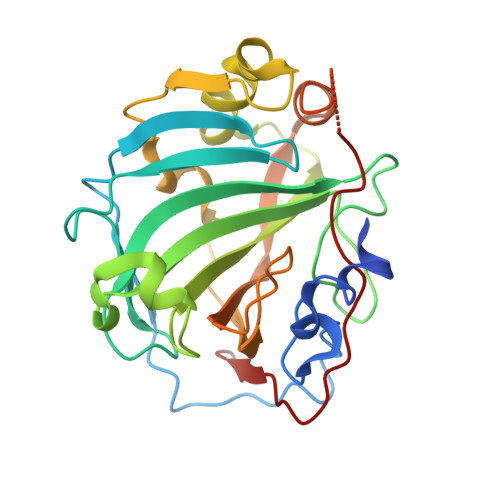
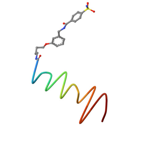
    The development of large synthetic ligands could be useful to target the sizeable surface areas involved in protein-protein interactions. Herein, we present long helical aromatic oligoamide foldamers bearing proteinogenic side chains that cover up to 450 Å 2 of the human carbonic anhydrase II (HCA) surface. The foldamers are composed of aminoquinolinecarboxylic acids bearing proteinogenic side chains and of more flexible aminomethyl-pyridinecarboxylic acids that enhance helix handedness dynamics. Crystal structures of HCA-foldamer complexes were obtained with a 9- and a 14-mer both showing extensive protein-foldamer hydrophobic contacts. In addition, foldamer-foldamer interactions seem to be prevalent in the crystal packing, leading to the peculiar formation of an HCA superhelix wound around a rod of stacked foldamers. Solution studies confirm the positioning of the foldamer at the protein surface as well as a dimerization of the complexes.


  • Organizational Affiliation

    CBMN (UMR5248), Univ. Bordeaux-CNRS-INP, Institut Européen de Chimie et Biologie, 2 rue Escarpit, 33600, Pessac, France.


Macromolecules
Find similar proteins by:  (by identity cutoff)  |  3D Structure
Entity ID: 1
MoleculeChains Sequence LengthOrganismDetailsImage
Carbonic anhydrase 2259Homo sapiensMutation(s): 0 
Gene Names: CA2
EC: 4.2.1.1 (PDB Primary Data), 4.2.1.69 (UniProt)
UniProt & NIH Common Fund Data Resources
Find proteins for P00918 (Homo sapiens)
Explore P00918 
Go to UniProtKB:  P00918
PHAROS:  P00918
GTEx:  ENSG00000104267 
Entity Groups  
Sequence Clusters30% Identity50% Identity70% Identity90% Identity95% Identity100% Identity
UniProt GroupP00918
Sequence Annotations
Expand
  • Reference Sequence

Find similar proteins by:  Sequence   |   3D Structure  

Entity ID: 2
MoleculeChains Sequence LengthOrganismDetailsImage
Aromatic foldamer16synthetic constructMutation(s): 0 
Sequence Annotations
Expand
  • Reference Sequence
Small Molecules
Ligands 1 Unique
IDChains Name / Formula / InChI Key2D Diagram3D Interactions
ZN
Query on ZN

Download Ideal Coordinates CCD File 
C [auth A]ZINC ION
Zn
PTFCDOFLOPIGGS-UHFFFAOYSA-N
Experimental Data & Validation

Experimental Data

  • Method: X-RAY DIFFRACTION
  • Resolution: 2.68 Å
  • R-Value Free: 0.260 
  • R-Value Work: 0.215 
  • R-Value Observed: 0.217 
  • Space Group: P 21 21 2
Unit Cell:
Length ( Å )Angle ( ˚ )
a = 79.3α = 90
b = 80.96β = 90
c = 45.62γ = 90
Software Package:
Software NamePurpose
REFMACrefinement
XDSdata reduction
XDSdata scaling
PHASERphasing

Structure Validation

View Full Validation Report


Entry History 

Deposition Data

Revision History  (Full details and data files)

  • Version 1.0: 2019-09-18
    Type: Initial release
  • Version 2.0: 2023-11-15
    Changes: Atomic model, Data collection, Database references, Derived calculations
  • Version 2.1: 2024-01-24
    Changes: Refinement description
  • Version 3.0: 2024-08-14
    Changes: Atomic model, Data collection, Database references, Derived calculations, Non-polymer description, Other, Polymer sequence, Refinement description, Source and taxonomy, Structure summary