6PPU

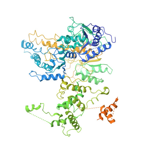
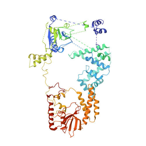
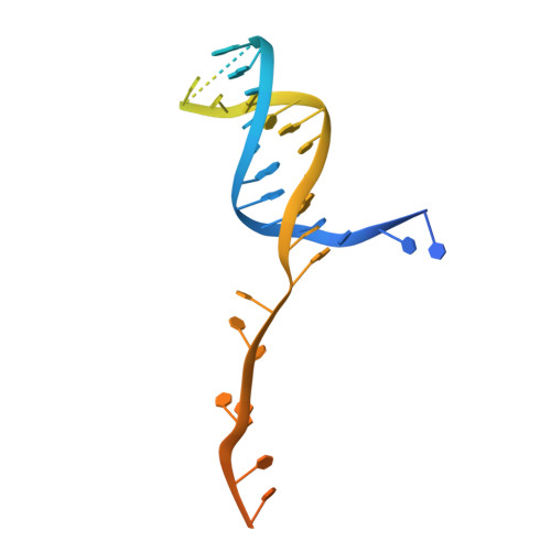
Cryo-EM structure of AdnAB-AMPPNP-DNA complex


Experimental Data Snapshot

  • Method: ELECTRON MICROSCOPY
  • Resolution: 3.50 Å
  • Aggregation State: PARTICLE 
  • Reconstruction Method: SINGLE PARTICLE 

wwPDB Validation   3D Report Full Report


This is version 1.3 of the entry. See complete history


Literature

Structures and single-molecule analysis of bacterial motor nuclease AdnAB illuminate the mechanism of DNA double-strand break resection.

Jia, N.Unciuleac, M.C.Xue, C.Greene, E.C.Patel, D.J.Shuman, S.

(2019) Proc Natl Acad Sci U S A 116: 24507-24516

  • DOI: https://doi.org/10.1073/pnas.1913546116
  • Primary Citation of Related Structures:  
    6PPJ, 6PPR, 6PPU

  • PubMed Abstract: 

    Mycobacterial AdnAB is a heterodimeric helicase-nuclease that initiates homologous recombination by resecting DNA double-strand breaks (DSBs). The AdnA and AdnB subunits are each composed of an N-terminal motor domain and a C-terminal nuclease domain. Here we report cryoelectron microscopy (cryo-EM) structures of AdnAB in three functional states: in the absence of DNA and in complex with forked duplex DNAs before and after cleavage of the 5' single-strand DNA (ssDNA) tail by the AdnA nuclease. The structures reveal the path of the 5' ssDNA through the AdnA nuclease domain and the mechanism of 5' strand cleavage; the path of the 3' tracking strand through the AdnB motor and the DNA contacts that couple ATP hydrolysis to mechanical work; the position of the AdnA iron-sulfur cluster subdomain at the Y junction and its likely role in maintaining the split trajectories of the unwound 5' and 3' strands. Single-molecule DNA curtain analysis of DSB resection reveals that AdnAB is highly processive but prone to spontaneous pausing at random sites on duplex DNA. A striking property of AdnAB is that the velocity of DSB resection slows after the enzyme experiences a spontaneous pause. Our results highlight shared as well as distinctive properties of AdnAB vis-à-vis the RecBCD and AddAB clades of bacterial DSB-resecting motor nucleases.


  • Organizational Affiliation

    Structural Biology Program, Sloan Kettering Institute, New York, NY 10065.


Macromolecules

Find similar proteins by:  (by identity cutoff)  |  3D Structure
Entity ID: 1
MoleculeChains Sequence LengthOrganismDetailsImage
UvrD/REP helicaseA [auth B]1,095Mycolicibacterium smegmatis MC2 155Mutation(s): 0 
Gene Names: MSMEI_1900
EC: 5.6.2.4
UniProt
Find proteins for I7FZ56 (Mycolicibacterium smegmatis (strain ATCC 700084 / mc(2)155))
Explore I7FZ56 
Go to UniProtKB:  I7FZ56
Entity Groups  
Sequence Clusters30% Identity50% Identity70% Identity90% Identity95% Identity100% Identity
UniProt GroupI7FZ56
Sequence Annotations
Expand
  • Reference Sequence
Find similar proteins by:  (by identity cutoff)  |  3D Structure
Entity ID: 2
MoleculeChains Sequence LengthOrganismDetailsImage
ATP-dependent DNA helicase (UvrD/REP)B [auth A]724Mycolicibacterium smegmatisMutation(s): 0 
Gene Names: pcrA_1ERS451418_01973
EC: 3.6.4.12 (PDB Primary Data), 5.6.2.4 (UniProt)
UniProt
Find proteins for A0QTR9 (Mycolicibacterium smegmatis (strain ATCC 700084 / mc(2)155))
Explore A0QTR9 
Go to UniProtKB:  A0QTR9
Entity Groups  
Sequence Clusters30% Identity50% Identity70% Identity90% Identity95% Identity100% Identity
UniProt GroupA0QTR9
Sequence Annotations
Expand
  • Reference Sequence
Find similar nucleic acids by:  (by identity cutoff)  |  3D Structure
Entity ID: 3
MoleculeChains LengthOrganismImage
DNA (29-MER)C [auth X]70Bacillus subtilis subsp. subtilis str. 168
Sequence Annotations
Expand
  • Reference Sequence
Experimental Data & Validation

Experimental Data

  • Method: ELECTRON MICROSCOPY
  • Resolution: 3.50 Å
  • Aggregation State: PARTICLE 
  • Reconstruction Method: SINGLE PARTICLE 
EM Software:
TaskSoftware PackageVersion
RECONSTRUCTIONRELION2.1

Structure Validation

View Full Validation Report


Entry History 

Deposition Data

Revision History  (Full details and data files)

  • Version 1.0: 2019-11-20
    Type: Initial release
  • Version 1.1: 2019-12-04
    Changes: Database references
  • Version 1.2: 2019-12-18
    Changes: Database references
  • Version 1.3: 2024-03-20
    Changes: Data collection, Database references