6OR0

Crystal structure of Insulin from Non-merohedrally twinned crystals


Experimental Data Snapshot

  • Method: X-RAY DIFFRACTION
  • Resolution: 1.55 Å
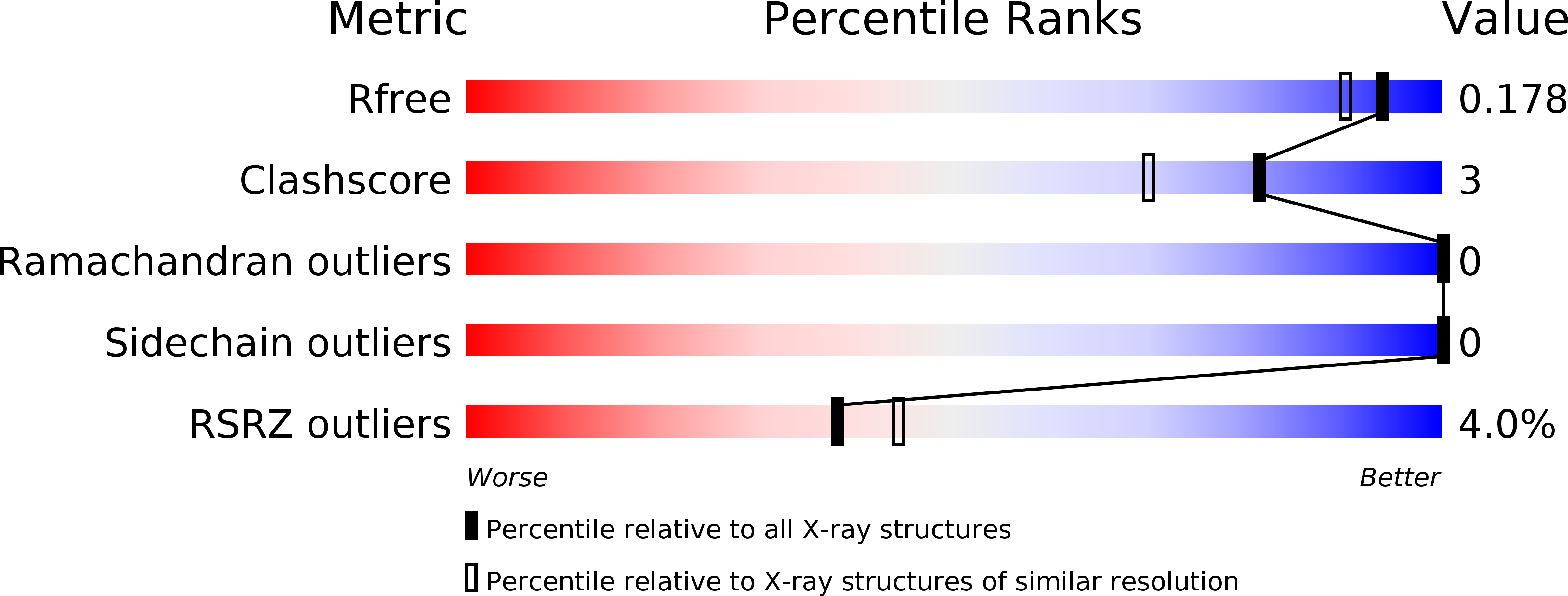
  • R-Value Free: 0.215 
  • R-Value Work: 0.166 
  • R-Value Observed: 0.166 

wwPDB Validation   3D Report Full Report


This is version 1.1 of the entry. See complete history


Literature

Non-merohedral twinning: from minerals to proteins.

Sevvana, M.Ruf, M.Uson, I.Sheldrick, G.M.Herbst-Irmer, R.

(2019) Acta Crystallogr D Struct Biol 75: 1040-1050

  • DOI: https://doi.org/10.1107/S2059798319010179
  • Primary Citation of Related Structures:  
    6OQZ, 6OR0

  • PubMed Abstract: 

    In contrast to twinning by merohedry, the reciprocal lattices of the different domains of non-merohedral twins do not overlap exactly. This leads to three kinds of reflections: reflections with no overlap, reflections with an exact overlap and reflections with a partial overlap of a reflection from a second domain. This complicates the unit-cell determination, indexing, data integration and scaling of X-ray diffraction data. However, with hindsight it is possible to detwin the data because there are reflections that are not affected by the twinning. In this article, the successful solution and refinement of one mineral, one organometallic and two protein non-merohedral twins using a common strategy are described. The unit-cell constants and the orientation matrices were determined by the program CELL_NOW. The data were then integrated with SAINT. TWINABS was used for scaling, empirical absorption corrections and the generation of two different data files, one with detwinned data for structure solution and refinement and a second one for (usually more accurate) structure refinement against total integrated intensities. The structures were solved by experimental phasing using SHELXT for the first two structures and SHELXC/D/E for the two protein structures; all models were refined with SHELXL.


  • Organizational Affiliation

    Department of Biological Sciences, Purdue University, West Lafayette, IN 47907, USA.


Macromolecules

Find similar proteins by:  Sequence   |   3D Structure  

Entity ID: 1
MoleculeChains Sequence LengthOrganismDetailsImage
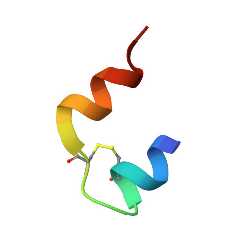
Insulin chain A21Bos taurusMutation(s): 0 
UniProt
Find proteins for P01317 (Bos taurus)
Explore P01317 
Go to UniProtKB:  P01317
Entity Groups  
Sequence Clusters30% Identity50% Identity70% Identity90% Identity95% Identity100% Identity
UniProt GroupP01317
Sequence Annotations
Expand
  • Reference Sequence
Find similar proteins by:  (by identity cutoff)  |  3D Structure
Entity ID: 2
MoleculeChains Sequence LengthOrganismDetailsImage
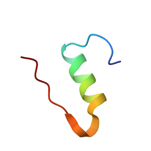
Insulin chain B29Bos taurusMutation(s): 0 
UniProt
Find proteins for P01317 (Bos taurus)
Explore P01317 
Go to UniProtKB:  P01317
Entity Groups  
Sequence Clusters30% Identity50% Identity70% Identity90% Identity95% Identity100% Identity
UniProt GroupP01317
Sequence Annotations
Expand
  • Reference Sequence
Experimental Data & Validation

Experimental Data

  • Method: X-RAY DIFFRACTION
  • Resolution: 1.55 Å
  • R-Value Free: 0.215 
  • R-Value Work: 0.166 
  • R-Value Observed: 0.166 
  • Space Group: I 21 3
Unit Cell:
Length ( Å )Angle ( ˚ )
a = 78.031α = 90
b = 78.031β = 90
c = 78.031γ = 90
Software Package:
Software NamePurpose
SHELXrefinement
SAINTdata reduction
SADABSdata scaling
SHELXDEphasing

Structure Validation

View Full Validation Report


Entry History & Funding Information

Deposition Data


Funding OrganizationLocationGrant Number
Spanish Ministry of Science, Innovation, and UniversitiesSpainBIO2015-64216-P
European Union (EU)European UnionMDM2014-0435

Revision History  (Full details and data files)

  • Version 1.0: 2019-12-11
    Type: Initial release
  • Version 1.1: 2024-10-23
    Changes: Data collection, Database references, Structure summary