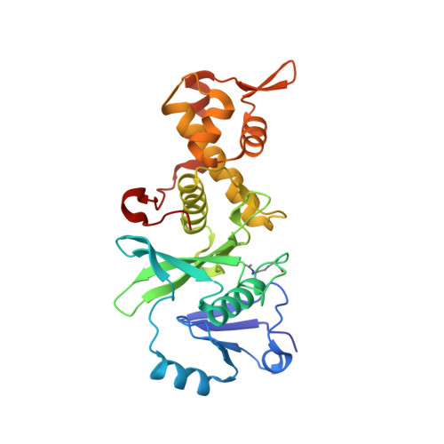
6OIQ

Crystal structure of MYST acetyltransferase domain in complex with inhibitor 63


Experimental Data Snapshot

  • Method: X-RAY DIFFRACTION
  • Resolution: 1.75 Å
  • R-Value Free: 0.227 
  • R-Value Work: 0.184 
  • R-Value Observed: 0.186 

wwPDB Validation   3D Report Full Report


Ligand Structure Quality Assessment 


This is version 1.3 of the entry. See complete history


Literature

Discovery of Benzoylsulfonohydrazides as Potent Inhibitors of the Histone Acetyltransferase KAT6A.

Leaver, D.J.Cleary, B.Nguyen, N.Priebbenow, D.L.Lagiakos, H.R.Sanchez, J.Xue, L.Huang, F.Sun, Y.Mujumdar, P.Mudududdla, R.Varghese, S.Teguh, S.Charman, S.A.White, K.L.Katneni, K.Cuellar, M.Strasser, J.M.Dahlin, J.L.Walters, M.A.Street, I.P.Monahan, B.J.Jarman, K.E.Sabroux, H.J.Falk, H.Chung, M.C.Hermans, S.J.Parker, M.W.Thomas, T.Baell, J.B.

(2019) J Med Chem 62: 7146-7159

  • DOI: https://doi.org/10.1021/acs.jmedchem.9b00665
  • Primary Citation of Related Structures:  
    6OIN, 6OIO, 6OIP, 6OIQ, 6OIR

  • PubMed Abstract: 

    A high-throughput screen for inhibitors of the histone acetyltransferase, KAT6A, led to identification of an aryl sulfonohydrazide derivative (CTX-0124143) that inhibited KAT6A with an IC 50 of 1.0 μM. Elaboration of the structure-activity relationship and medicinal chemistry optimization led to the discovery of WM-8014 ( 97 ), a highly potent inhibitor of KAT6A (IC 50 = 0.008 μM). WM-8014 competes with acetyl-CoA (Ac-CoA), and X-ray crystallographic analysis demonstrated binding to the Ac-CoA binding site. Through inhibition of KAT6A activity, WM-8014 induces cellular senescence and represents a unique pharmacological tool.


  • Organizational Affiliation

    Cancer Therapeutics CRC , 343 Royal Parade , Parkville , Victoria 3052 , Australia.


Macromolecules
Find similar proteins by:  (by identity cutoff)  |  3D Structure
Entity ID: 1
MoleculeChains Sequence LengthOrganismDetailsImage
Histone acetyltransferase KAT8273Homo sapiensMutation(s): 4 
Gene Names: KAT8MOFMYST1PP7073
EC: 2.3.1.48 (PDB Primary Data), 2.3.1 (UniProt)
UniProt & NIH Common Fund Data Resources
Find proteins for Q9H7Z6 (Homo sapiens)
Explore Q9H7Z6 
Go to UniProtKB:  Q9H7Z6
PHAROS:  Q9H7Z6
GTEx:  ENSG00000103510 
Entity Groups  
Sequence Clusters30% Identity50% Identity70% Identity90% Identity95% Identity100% Identity
UniProt GroupQ9H7Z6
Sequence Annotations
Expand
  • Reference Sequence
Small Molecules
Ligands 3 Unique
IDChains Name / Formula / InChI Key2D Diagram3D Interactions
MLV
Query on MLV

Download Ideal Coordinates CCD File 
C [auth A]2-fluoro-N'-(phenylsulfonyl)[1,1'-biphenyl]-3-carbohydrazide
C19 H15 F N2 O3 S
DUTMMJVUYVOSEW-UHFFFAOYSA-N
GOL
Query on GOL

Download Ideal Coordinates CCD File 
D [auth A]GLYCEROL
C3 H8 O3
PEDCQBHIVMGVHV-UHFFFAOYSA-N
ZN
Query on ZN

Download Ideal Coordinates CCD File 
B [auth A]ZINC ION
Zn
PTFCDOFLOPIGGS-UHFFFAOYSA-N
Modified Residues  1 Unique
IDChains TypeFormula2D DiagramParent
ALY
Query on ALY
A
L-PEPTIDE LINKINGC8 H16 N2 O3LYS
Experimental Data & Validation

Experimental Data

  • Method: X-RAY DIFFRACTION
  • Resolution: 1.75 Å
  • R-Value Free: 0.227 
  • R-Value Work: 0.184 
  • R-Value Observed: 0.186 
  • Space Group: P 21 21 21
Unit Cell:
Length ( Å )Angle ( ˚ )
a = 46.603α = 90
b = 56.772β = 90
c = 122.277γ = 90
Software Package:
Software NamePurpose
REFMACrefinement
MOSFLMdata reduction
SCALAdata scaling
PHASERphasing

Structure Validation

View Full Validation Report


Ligand Structure Quality Assessment 


Entry History 

Deposition Data

Revision History  (Full details and data files)

  • Version 1.0: 2019-07-03
    Type: Initial release
  • Version 1.1: 2019-07-17
    Changes: Data collection, Database references
  • Version 1.2: 2019-08-21
    Changes: Data collection, Database references
  • Version 1.3: 2024-10-23
    Changes: Data collection, Database references, Derived calculations, Structure summary