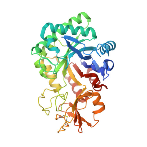
Crystal structure of CsChiL, a chitinase from Chitiniphilus shinanonensis
Ueda, M., Sonoda, N., Shimosaka, M., Arai, R.To be published.
Experimental Data Snapshot
Starting Model: experimental
View more details
Entity ID: 1 | |||||
|---|---|---|---|---|---|
| Molecule | Chains | Sequence Length | Organism | Details | Image |
| Family 18 chitinase | 372 | Chitiniphilus shinanonensis | Mutation(s): 0 Gene Names: chiL |  | |
UniProt | |||||
Find proteins for F8WSX2 (Chitiniphilus shinanonensis) Explore F8WSX2 Go to UniProtKB: F8WSX2 | |||||
Entity Groups | |||||
| Sequence Clusters | 30% Identity50% Identity70% Identity90% Identity95% Identity100% Identity | ||||
| UniProt Group | F8WSX2 | ||||
Sequence AnnotationsExpand | |||||
| |||||
| Ligands 7 Unique | |||||
|---|---|---|---|---|---|
| ID | Chains | Name / Formula / InChI Key | 2D Diagram | 3D Interactions | |
| NAG Query on NAG | N [auth A], X [auth B] | 2-acetamido-2-deoxy-beta-D-glucopyranose C8 H15 N O6 OVRNDRQMDRJTHS-FMDGEEDCSA-N |  | ||
| PG4 Query on PG4 | R [auth B], S [auth B], T [auth B] | TETRAETHYLENE GLYCOL C8 H18 O5 UWHCKJMYHZGTIT-UHFFFAOYSA-N |  | ||
| PGE Query on PGE | J [auth A], U [auth B] | TRIETHYLENE GLYCOL C6 H14 O4 ZIBGPFATKBEMQZ-UHFFFAOYSA-N |  | ||
| MLI Query on MLI | G [auth A], H [auth A], I [auth A], P [auth B], Q [auth B] | MALONATE ION C3 H2 O4 OFOBLEOULBTSOW-UHFFFAOYSA-L |  | ||
| MXE Query on MXE | F [auth A] | 2-METHOXYETHANOL C3 H8 O2 XNWFRZJHXBZDAG-UHFFFAOYSA-N |  | ||
| EDO Query on EDO | K [auth A], L [auth A], M [auth A], V [auth B], W [auth B] | 1,2-ETHANEDIOL C2 H6 O2 LYCAIKOWRPUZTN-UHFFFAOYSA-N |  | ||
| ACT Query on ACT | E [auth A], O [auth B] | ACETATE ION C2 H3 O2 QTBSBXVTEAMEQO-UHFFFAOYSA-M |  | ||
| Length ( Å ) | Angle ( ˚ ) |
|---|---|
| a = 68.982 | α = 90 |
| b = 82.502 | β = 90 |
| c = 131.818 | γ = 90 |
| Software Name | Purpose |
|---|---|
| REFMAC | refinement |
| HKL-2000 | data scaling |
| MOLREP | phasing |
| PDB_EXTRACT | data extraction |
| HKL-2000 | data reduction |
| Funding Organization | Location | Grant Number |
|---|---|---|
| Japan Society for the Promotion of Science | Japan | JP24780097 |
| Japan Society for the Promotion of Science | Japan | JP24580107 |
| Japan Society for the Promotion of Science | Japan | JP24113707 |
| Japan Society for the Promotion of Science | Japan | JP16K05841 |
| Japan Society for the Promotion of Science | Japan | JP16H00761 |
| Japan Society for the Promotion of Science | Japan | JP17KK0104 |
| Japan Society for the Promotion of Science | Japan | JP19H02522 |