6KDM

HIV-1 reverse transcriptase with Q151M/Y115F/F116Y:DNA:entecavir 5'-triphosphate ternary complex


Experimental Data Snapshot

  • Method: X-RAY DIFFRACTION
  • Resolution: 2.32 Å
  • R-Value Free: 0.223 
  • R-Value Work: 0.190 
  • R-Value Observed: 0.192 

Starting Model: experimental
View more details

wwPDB Validation   3D Report Full Report


Ligand Structure Quality Assessment 


This is version 1.1 of the entry. See complete history


Literature

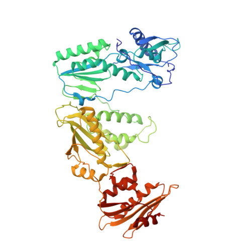
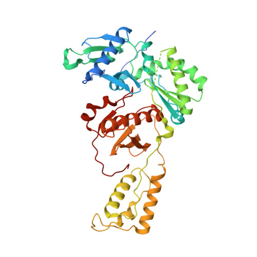
Structural features in common of HBV and HIV-1 resistance against chirally-distinct nucleoside analogues entecavir and lamivudine.

Yasutake, Y.Hattori, S.I.Tamura, N.Matsuda, K.Kohgo, S.Maeda, K.Mitsuya, H.

(2020) Sci Rep 10: 3021-3021

  • DOI: https://doi.org/10.1038/s41598-020-59775-w
  • Primary Citation of Related Structures:  
    6KDJ, 6KDK, 6KDM, 6KDN, 6KDO

  • PubMed Abstract: 

    Chronic hepatitis B virus (HBV) infection is a major public health problem that affects millions of people worldwide. Nucleoside analogue reverse transcriptase (RT) inhibitors, such as entecavir (ETV) and lamivudine (3TC), serve as crucial anti-HBV drugs. However, structural studies of HBV RT have been hampered due to its unexpectedly poor solubility. Here, we show that human immunodeficiency virus type-1 (HIV-1) with HBV-associated amino acid substitutions Y115F/F116Y/Q151M in its RT (HIV Y115F/F116Y/Q151M ) is highly susceptible to ETV and 3TC. Additionally, we experimentally simulated previously reported ETV/3TC resistance for HBV using HIV Y115F/F116Y/Q151M with F160M/M184V (L180M/M204V in HBV RT) substituted. We determined crystal structures for HIV-1 RT Y115F/F116Y/Q151M :DNA complexed with 3TC-triphosphate (3TC-TP)/ETV-triphosphate (ETV-TP)/dCTP/dGTP. These structures revealed an atypically tight binding conformation of 3TC-TP, where the Met184 side-chain is pushed away by the oxathiolane of 3TC-TP and exocyclic methylene of ETV-TP. Structural analysis of RT Y115F/F116Y/Q151M/F160M/M184V :DNA:3TC-TP also demonstrated that the loosely bound 3TC-TP is misaligned at the active site to prevent a steric clash with the side chain γ-methyl of Val184. These findings shed light on the common structural mechanism of HBV and HIV-1 resistance to 3TC and ETV and should aid in the design of new agents to overcome drug resistance to 3TC and ETV.


  • Organizational Affiliation

    Bioproduction Research Institute, National Institute of Advanced Industrial Science and Technology (AIST), Sapporo, 062-8517, Japan. y-yasutake@aist.go.jp.


Macromolecules

Find similar proteins by:  (by identity cutoff)  |  3D Structure
Entity ID: 1
MoleculeChains Sequence LengthOrganismDetailsImage
HIV-1 reverse transcriptase p66 subunitA,
D [auth C]
557Human immunodeficiency virus 1Mutation(s): 5 
Gene Names: pol
UniProt
Find proteins for P12497 (Human immunodeficiency virus type 1 group M subtype B (isolate NY5))
Explore P12497 
Go to UniProtKB:  P12497
Entity Groups  
Sequence Clusters30% Identity50% Identity70% Identity90% Identity95% Identity100% Identity
UniProt GroupP12497
Sequence Annotations
Expand
  • Reference Sequence
Find similar proteins by:  (by identity cutoff)  |  3D Structure
Entity ID: 2
MoleculeChains Sequence LengthOrganismDetailsImage
HIV-1 RT p51 subunitB,
E [auth D]
444Human immunodeficiency virus 1Mutation(s): 2 
UniProt
Find proteins for P12497 (Human immunodeficiency virus type 1 group M subtype B (isolate NY5))
Explore P12497 
Go to UniProtKB:  P12497
Entity Groups  
Sequence Clusters30% Identity50% Identity70% Identity90% Identity95% Identity100% Identity
UniProt GroupP12497
Sequence Annotations
Expand
  • Reference Sequence
Find similar nucleic acids by:  (by identity cutoff)  |  3D Structure
Entity ID: 3
MoleculeChains LengthOrganismImage
DNA/RNA (38-MER)C [auth E],
F
38synthetic construct
Sequence Annotations
Expand
  • Reference Sequence
Small Molecules
Ligands 3 Unique
IDChains Name / Formula / InChI Key2D Diagram3D Interactions
ET9 (Subject of Investigation/LOI)
Query on ET9

Download Ideal Coordinates CCD File 
I [auth A],
M [auth C]
[[(1R,3S,5S)-3-(2-azanyl-6-oxidanylidene-3H-purin-9-yl)-2-methylidene-5-oxidanyl-cyclopentyl]methoxy-oxidanyl-phosphory l] phosphono hydrogen phosphate
C12 H18 N5 O12 P3
YMBBDUCQYPKKJK-FXQIFTODSA-N
GOL
Query on GOL

Download Ideal Coordinates CCD File 
G [auth A]
J [auth B]
K [auth E]
N [auth D]
O [auth D]
G [auth A],
J [auth B],
K [auth E],
N [auth D],
O [auth D],
P [auth D]
GLYCEROL
C3 H8 O3
PEDCQBHIVMGVHV-UHFFFAOYSA-N
MG
Query on MG

Download Ideal Coordinates CCD File 
H [auth A],
L [auth C]
MAGNESIUM ION
Mg
JLVVSXFLKOJNIY-UHFFFAOYSA-N
Experimental Data & Validation

Experimental Data

  • Method: X-RAY DIFFRACTION
  • Resolution: 2.32 Å
  • R-Value Free: 0.223 
  • R-Value Work: 0.190 
  • R-Value Observed: 0.192 
  • Space Group: H 3
Unit Cell:
Length ( Å )Angle ( ˚ )
a = 284.447α = 90
b = 284.447β = 90
c = 95.773γ = 120
Software Package:
Software NamePurpose
PHENIXrefinement
XDSdata reduction
Aimlessdata scaling
PHENIXphasing

Structure Validation

View Full Validation Report


Ligand Structure Quality Assessment 


Entry History & Funding Information

Deposition Data


Funding OrganizationLocationGrant Number
Japan Agency for Medical Research and Development (AMED)JapanJP19fk0310113

Revision History  (Full details and data files)

  • Version 1.0: 2020-03-04
    Type: Initial release
  • Version 1.1: 2023-11-22
    Changes: Data collection, Database references, Derived calculations, Refinement description, Structure summary