6K2O

Structural basis of glycan recognition in globally predominant human P[8] rotavirus


Experimental Data Snapshot

  • Method: X-RAY DIFFRACTION
  • Resolution: 2.30 Å
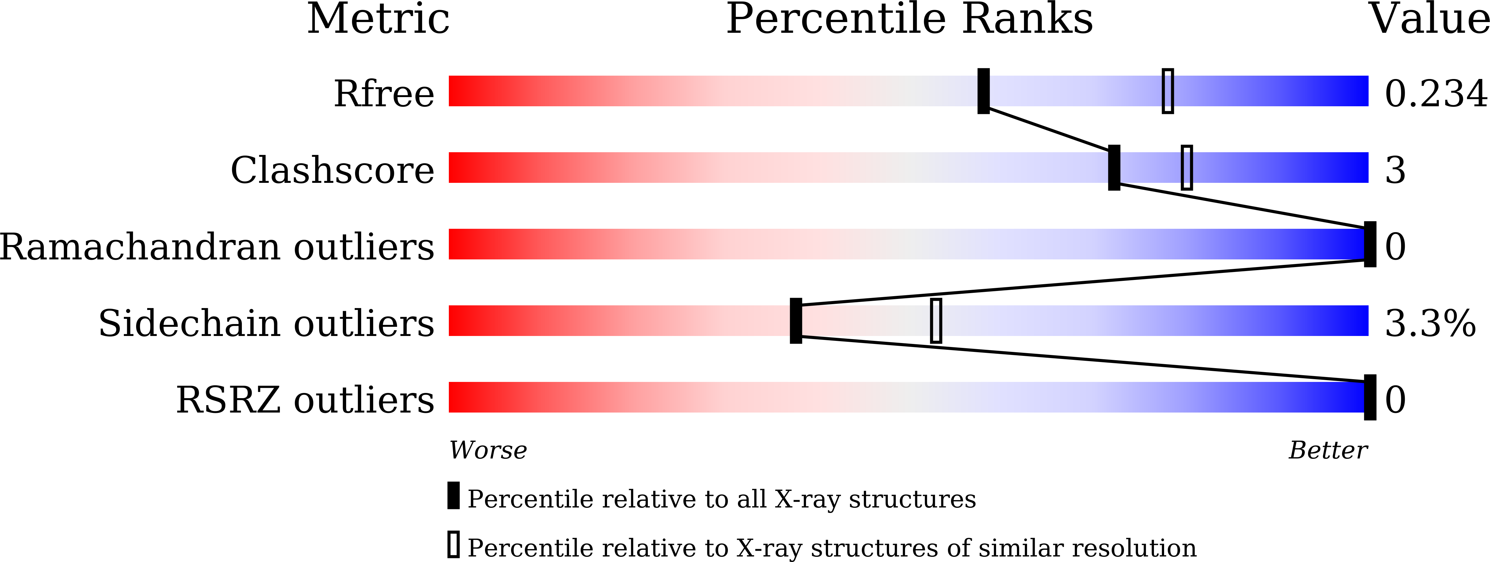
  • R-Value Free: 0.233 
  • R-Value Work: 0.203 
  • R-Value Observed: 0.205 

Starting Model: experimental
View more details

wwPDB Validation   3D Report Full Report


This is version 2.1 of the entry. See complete history


Literature

Structural Basis of Glycan Recognition in Globally Predominant Human P[8] Rotavirus.

Sun, X.Dang, L.Li, D.Qi, J.Wang, M.Chai, W.Zhang, Q.Wang, H.Bai, R.Tan, M.Duan, Z.

(2020) Virol Sin 35: 156-170

  • DOI: https://doi.org/10.1007/s12250-019-00164-7
  • Primary Citation of Related Structures:  
    6K2N, 6K2O

  • PubMed Abstract: 

    Rotavirus (RV) causes acute gastroenteritis in infants and children worldwide. Recent studies showed that glycans such as histo-blood group antigens (HBGAs) function as cell attachment factors affecting RV host susceptibility and prevalence. P[8] is the predominant RV genotype in humans, but the structural basis of how P[8] RVs interact with glycan ligands remains elusive. In this study, we characterized the interactions between P[8] VP8*s and glycans which showed that VP8*, the RV glycan binding domain, recognized both mucin core 2 and H type 1 antigens according to the ELISA-based oligosaccharide binding assays. Importantly, we determined the structural basis of P[8] RV-glycans interaction from the crystal structures of a Rotateq P[8] VP8* in complex with core 2 and H type 1 glycans at 1.8 Å and 2.3 Å, respectively, revealing a common binding pocket and similar binding mode. Structural and sequence analysis demonstrated that the glycan binding site is conserved among RVs in the P[II] genogroup, while genotype-specific amino acid variations determined different glycan binding preference. Our data elucidated the detailed structural basis of the interactions between human P[8] RVs and different host glycan factors, shedding light on RV infection, epidemiology, and development of anti-viral agents.


  • Organizational Affiliation

    National Health Commission Key Laboratory for Medical Virology and Viral Diseases, Beijing, 102206, China.


Macromolecules
Find similar proteins by:  (by identity cutoff)  |  3D Structure
Entity ID: 1
MoleculeChains Sequence LengthOrganismDetailsImage
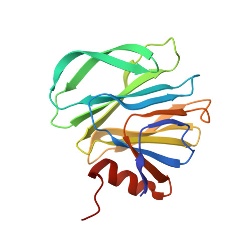
Outer capsid protein VP4163Rotavirus AMutation(s): 0 
UniProt
Find proteins for E2EA82 (Rotavirus A)
Explore E2EA82 
Go to UniProtKB:  E2EA82
Entity Groups  
Sequence Clusters30% Identity50% Identity70% Identity90% Identity95% Identity100% Identity
UniProt GroupE2EA82
Sequence Annotations
Expand
  • Reference Sequence
Oligosaccharides

Help

Entity ID: 2
MoleculeChains Length2D Diagram Glycosylation3D Interactions
alpha-L-fucopyranose-(1-2)-beta-D-galactopyranose-(1-3)-2-acetamido-2-deoxy-beta-D-glucopyranose-(1-3)-beta-D-galactopyranose
B
4N/A
Glycosylation Resources
GlyTouCan:  G00294TN
GlyCosmos:  G00294TN
GlyGen:  G00294TN
Small Molecules
Experimental Data & Validation

Experimental Data

  • Method: X-RAY DIFFRACTION
  • Resolution: 2.30 Å
  • R-Value Free: 0.233 
  • R-Value Work: 0.203 
  • R-Value Observed: 0.205 
  • Space Group: P 63 2 2
Unit Cell:
Length ( Å )Angle ( ˚ )
a = 80.454α = 90
b = 80.454β = 90
c = 116.593γ = 120
Software Package:
Software NamePurpose
HKL-2000data reduction
HKL-2000data scaling
PHENIXrefinement
PDB_EXTRACTdata extraction
PHASERphasing

Structure Validation

View Full Validation Report


Entry History & Funding Information

Deposition Data


Funding OrganizationLocationGrant Number
National Science Foundation (China)Chinano.2018ZA10711-001 and no.2017ZX10104001

Revision History  (Full details and data files)

  • Version 1.0: 2019-10-09
    Type: Initial release
  • Version 1.1: 2019-10-30
    Changes: Data collection, Database references
  • Version 1.2: 2020-05-13
    Changes: Data collection, Database references
  • Version 2.0: 2020-07-29
    Type: Remediation
    Reason: Carbohydrate remediation
    Changes: Advisory, Atomic model, Data collection, Derived calculations, Structure summary
  • Version 2.1: 2023-11-22
    Changes: Data collection, Database references, Derived calculations, Refinement description, Structure summary