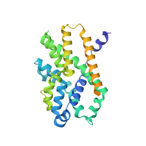
6IZ3

Structural basis for activity of TRIC counter-ion channels in calcium release


Experimental Data Snapshot

  • Method: X-RAY DIFFRACTION
  • Resolution: 3.79 Å
  • R-Value Free: 0.341 
  • R-Value Work: 0.299 
  • R-Value Observed: 0.301 

Starting Model: experimental
View more details

wwPDB Validation   3D Report Full Report


This is version 1.2 of the entry. See complete history


Literature

Structural basis for activity of TRIC counter-ion channels in calcium release.

Wang, X.H.Su, M.Gao, F.Xie, W.Zeng, Y.Li, D.L.Liu, X.L.Zhao, H.Qin, L.Li, F.Liu, Q.Clarke, O.B.Lam, S.M.Shui, G.H.Hendrickson, W.A.Chen, Y.H.

(2019) Proc Natl Acad Sci U S A 116: 4238-4243

  • DOI: https://doi.org/10.1073/pnas.1817271116
  • Primary Citation of Related Structures:  
    6IYU, 6IYX, 6IYZ, 6IZ0, 6IZ1, 6IZ3, 6IZ4, 6IZ5, 6IZ6, 6IZF

  • PubMed Abstract: 

    Trimeric intracellular cation (TRIC) channels are thought to provide counter-ion currents that facilitate the active release of Ca 2+ from intracellular stores. TRIC activity is controlled by voltage and Ca 2+ modulation, but underlying mechanisms have remained unknown. Here we describe high-resolution crystal structures of vertebrate TRIC-A and TRIC-B channels, both in Ca 2+ -bound and Ca 2+ -free states, and we analyze conductance properties in structure-inspired mutagenesis experiments. The TRIC channels are symmetric trimers, wherein we find a pore in each protomer that is gated by a highly conserved lysine residue. In the resting state, Ca 2+ binding at the luminal surface of TRIC-A, on its threefold axis, stabilizes lysine blockage of the pores. During active Ca 2+ release, luminal Ca 2+ depletion removes inhibition to permit the lysine-bearing and voltage-sensing helix to move in response to consequent membrane hyperpolarization. Diacylglycerol is found at interprotomer interfaces, suggesting a role in metabolic control.


  • Organizational Affiliation

    State Key Laboratory of Molecular Developmental Biology, Institute of Genetics and Developmental Biology, Chinese Academy of Sciences, Beijing 100101, China.


Macromolecules
Find similar proteins by:  (by identity cutoff)  |  3D Structure
Entity ID: 1
MoleculeChains Sequence LengthOrganismDetailsImage
Trimeric intracellular cation channel type B-B
A, B, C, D
314Xenopus laevisMutation(s): 0 
Gene Names: tmem38b-b
Membrane Entity: Yes 
UniProt
Find proteins for Q6GN30 (Xenopus laevis)
Explore Q6GN30 
Go to UniProtKB:  Q6GN30
Entity Groups  
Sequence Clusters30% Identity50% Identity70% Identity90% Identity95% Identity100% Identity
UniProt GroupQ6GN30
Sequence Annotations
Expand
  • Reference Sequence
Experimental Data & Validation

Experimental Data

  • Method: X-RAY DIFFRACTION
  • Resolution: 3.79 Å
  • R-Value Free: 0.341 
  • R-Value Work: 0.299 
  • R-Value Observed: 0.301 
  • Space Group: P 3 2 1
Unit Cell:
Length ( Å )Angle ( ˚ )
a = 190.124α = 90
b = 190.124β = 90
c = 117.587γ = 120
Software Package:
Software NamePurpose
PHENIXrefinement
Aimlessdata scaling
PDB_EXTRACTdata extraction
XDSdata reduction
PHENIXphasing

Structure Validation

View Full Validation Report


Entry History & Funding Information

Deposition Data


Funding OrganizationLocationGrant Number
Ministry of Science and Technology (China)China2016YFA0500503
Ministry of Science and Technology (China)China2015CB910102

Revision History  (Full details and data files)

  • Version 1.0: 2019-05-01
    Type: Initial release
  • Version 1.1: 2019-11-06
    Changes: Data collection, Database references
  • Version 1.2: 2023-11-22
    Changes: Data collection, Database references, Refinement description