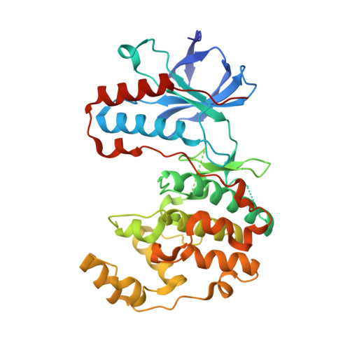
6HWU

Crystal structure of p38alpha in complex with a photoswitchable 2-Azothiazol-based Inhibitor (compound 2)


Experimental Data Snapshot

  • Method: X-RAY DIFFRACTION
  • Resolution: 2.30 Å
  • R-Value Free: 0.247 
  • R-Value Work: 0.191 
  • R-Value Observed: 0.193 

Starting Model: experimental
View more details

wwPDB Validation   3D Report Full Report


Ligand Structure Quality Assessment 


This is version 1.3 of the entry. See complete history


Literature

2-Azo-, 2-diazocine-thiazols and 2-azo-imidazoles as photoswitchable kinase inhibitors: limitations and pitfalls of the photoswitchable inhibitor approach.

Schehr, M.Ianes, C.Weisner, J.Heintze, L.Muller, M.P.Pichlo, C.Charl, J.Brunstein, E.Ewert, J.Lehr, M.Baumann, U.Rauh, D.Knippschild, U.Peifer, C.Herges, R.

(2019) Photochem Photobiol Sci 18: 1398-1407

  • DOI: https://doi.org/10.1039/c9pp00010k
  • Primary Citation of Related Structures:  
    6HMP, 6HMR, 6HWT, 6HWU, 6HWV

  • PubMed Abstract: 

    In photopharmacology, photoswitchable compounds including azobenzene or other diarylazo moieties exhibit bioactivity against a target protein typically in the slender E-configuration, whereas the rather bulky Z-configuration usually is pharmacologically less potent. Herein we report the design, synthesis and photochemical/inhibitory characterization of new photoswitchable kinase inhibitors targeting p38α MAPK and CK1δ. A well characterized inhibitor scaffold was used to attach arylazo- and diazocine moieties. When the isolated isomers, or the photostationary state (PSS) of isomers, were tested in commonly used in vitro kinase assays, however, only small differences in activity were observed. X-ray analyses of ligand-bound p38α MAPK and CK1δ complexes revealed dynamic conformational adaptations of the protein with respect to both isomers. More importantly, irreversible reduction of the azo group to the corresponding hydrazine was observed. Independent experiments revealed that reducing agents such as DTT (dithiothreitol) and GSH (glutathione) that are typically used for protein stabilization in biological assays were responsible. Two further sources of error are the concentration dependence of the E-Z-switching efficiency and artefacts due to incomplete exclusion of light during testing. Our findings may also apply to a number of previously investigated azobenzene-based photoswitchable inhibitors.


  • Organizational Affiliation

    Otto Diels-Institute of Organic Chemistry, Christian Albrechts University Kiel, Otto-Hahn-Platz 4, 24118 Kiel, Germany. rherges@oc.uni-kiel.de.


Macromolecules
Find similar proteins by:  (by identity cutoff)  |  3D Structure
Entity ID: 1
MoleculeChains Sequence LengthOrganismDetailsImage
Mitogen-activated protein kinase 14362Homo sapiensMutation(s): 0 
Gene Names: MAPK14CSBPCSBP1CSBP2CSPB1MXI2SAPK2A
EC: 2.7.11.24
UniProt & NIH Common Fund Data Resources
Find proteins for Q16539 (Homo sapiens)
Explore Q16539 
Go to UniProtKB:  Q16539
PHAROS:  Q16539
GTEx:  ENSG00000112062 
Entity Groups  
Sequence Clusters30% Identity50% Identity70% Identity90% Identity95% Identity100% Identity
UniProt GroupQ16539
Sequence Annotations
Expand
  • Reference Sequence
Small Molecules
Ligands 2 Unique
IDChains Name / Formula / InChI Key2D Diagram3D Interactions
GE5 (Subject of Investigation/LOI)
Query on GE5

Download Ideal Coordinates CCD File 
E [auth A]3-(2,5-dimethoxyphenyl)-~{N}-[4-[4-(4-fluorophenyl)-2-[(~{E})-phenyldiazenyl]-1,3-thiazol-5-yl]pyridin-2-yl]propanamide
C31 H26 F N5 O3 S
OCIGBTIRVZOQBN-BSRQYYOTSA-N
BOG
Query on BOG

Download Ideal Coordinates CCD File 
B [auth A],
C [auth A],
D [auth A]
octyl beta-D-glucopyranoside
C14 H28 O6
HEGSGKPQLMEBJL-RKQHYHRCSA-N
Experimental Data & Validation

Experimental Data

  • Method: X-RAY DIFFRACTION
  • Resolution: 2.30 Å
  • R-Value Free: 0.247 
  • R-Value Work: 0.191 
  • R-Value Observed: 0.193 
  • Space Group: P 21 21 21
Unit Cell:
Length ( Å )Angle ( ˚ )
a = 66.67α = 90
b = 74.69β = 90
c = 77.24γ = 90
Software Package:
Software NamePurpose
PHENIXrefinement
XDSdata reduction
XSCALEdata scaling
PHASERphasing

Structure Validation

View Full Validation Report


Ligand Structure Quality Assessment 


Entry History & Funding Information

Deposition Data


Funding OrganizationLocationGrant Number
German Federal Ministry for Education and ResearchGermanyBMBF 01GS08104
German Federal Ministry for Education and ResearchGermany01ZX1303C

Revision History  (Full details and data files)

  • Version 1.0: 2019-04-17
    Type: Initial release
  • Version 1.1: 2019-06-19
    Changes: Data collection, Database references
  • Version 1.2: 2020-07-29
    Type: Remediation
    Reason: Carbohydrate remediation
    Changes: Data collection, Derived calculations, Structure summary
  • Version 1.3: 2024-01-24
    Changes: Data collection, Database references, Refinement description, Structure summary