6HQU

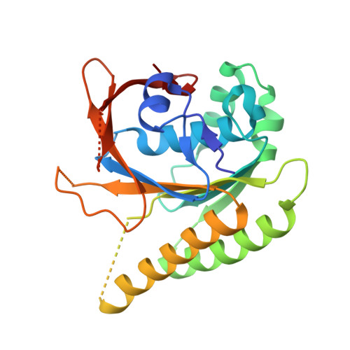
Humanised RadA mutant HumRadA22 in complex with a recombined BRC repeat 8-2


Experimental Data Snapshot

  • Method: X-RAY DIFFRACTION
  • Resolution: 1.97 Å
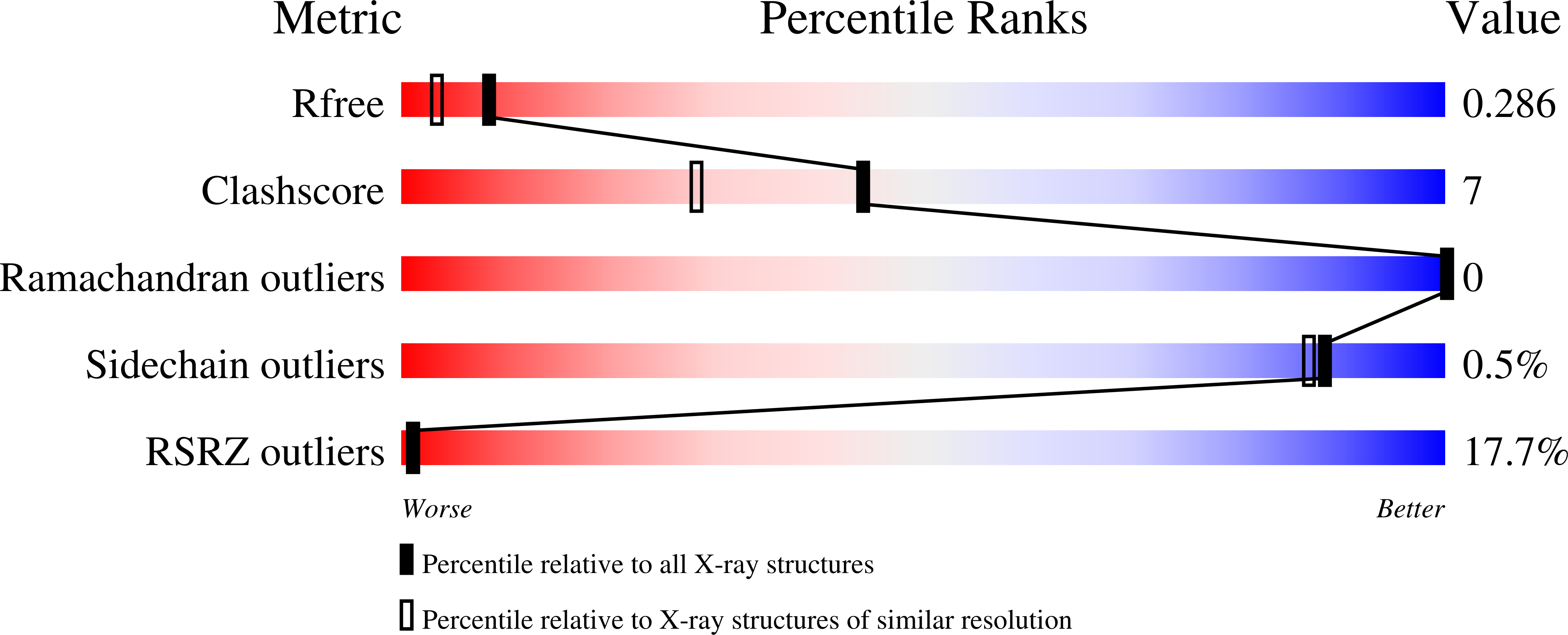
  • R-Value Free: 0.271 
  • R-Value Work: 0.263 
  • R-Value Observed: 0.263 

Starting Model: experimental
View more details

wwPDB Validation   3D Report Full Report


Ligand Structure Quality Assessment 


This is version 2.2 of the entry. See complete history


Literature

Improved RAD51 binders through motif shuffling based on the modularity of BRC repeats.

Lindenburg, L.H.Pantelejevs, T.Gielen, F.Zuazua-Villar, P.Butz, M.Rees, E.Kaminski, C.F.Downs, J.A.Hyvonen, M.Hollfelder, F.

(2021) Proc Natl Acad Sci U S A 118

  • DOI: https://doi.org/10.1073/pnas.2017708118
  • Primary Citation of Related Structures:  
    6HQU

  • PubMed Abstract: 

    Exchanges of protein sequence modules support leaps in function unavailable through point mutations during evolution. Here we study the role of the two RAD51-interacting modules within the eight binding BRC repeats of BRCA2. We created 64 chimeric repeats by shuffling these modules and measured their binding to RAD51. We found that certain shuffled module combinations were stronger binders than any of the module combinations in the natural repeats. Surprisingly, the contribution from the two modules was poorly correlated with affinities of natural repeats, with a weak BRC8 repeat containing the most effective N-terminal module. The binding of the strongest chimera, BRC8-2, to RAD51 was improved by -2.4 kCal/mol compared to the strongest natural repeat, BRC4. A crystal structure of RAD51:BRC8-2 complex shows an improved interface fit and an extended β-hairpin in this repeat. BRC8-2 was shown to function in human cells, preventing the formation of nuclear RAD51 foci after ionizing radiation.


  • Organizational Affiliation

    Department of Biochemistry, University of Cambridge, Cambridge CB2 1GA, United Kingdom.


Macromolecules
Find similar proteins by:  (by identity cutoff)  |  3D Structure
Entity ID: 1
MoleculeChains Sequence LengthOrganismDetailsImage
DNA repair and recombination protein RadA
A, B, C, D, E
A, B, C, D, E, F, G, H
231Pyrococcus furiosus DSM 3638Mutation(s): 0 
Gene Names: radAPF1926
Entity Groups  
Sequence Clusters30% Identity50% Identity70% Identity90% Identity95% Identity100% Identity
Sequence Annotations
Expand
  • Reference Sequence
Find similar proteins by:  (by identity cutoff)  |  3D Structure
Entity ID: 2
MoleculeChains Sequence LengthOrganismDetailsImage
Breast cancer type 2 susceptibility
I, J, K, L, M
I, J, K, L, M, N, O
38Homo sapiensMutation(s): 0 
UniProt & NIH Common Fund Data Resources
Find proteins for P51587 (Homo sapiens)
Explore P51587 
Go to UniProtKB:  P51587
PHAROS:  P51587
GTEx:  ENSG00000139618 
Entity Groups  
Sequence Clusters30% Identity50% Identity70% Identity90% Identity95% Identity100% Identity
UniProt GroupP51587
Sequence Annotations
Expand
  • Reference Sequence
Small Molecules
Ligands 2 Unique
IDChains Name / Formula / InChI Key2D Diagram3D Interactions
ADP
Query on ADP

Download Ideal Coordinates CCD File 
BA [auth G]
DA [auth H]
P [auth A]
R [auth B]
T [auth C]
BA [auth G],
DA [auth H],
P [auth A],
R [auth B],
T [auth C],
V [auth D],
X [auth E],
Z [auth F]
ADENOSINE-5'-DIPHOSPHATE
C10 H15 N5 O10 P2
XTWYTFMLZFPYCI-KQYNXXCUSA-N
MG
Query on MG

Download Ideal Coordinates CCD File 
AA [auth F]
CA [auth G]
EA [auth H]
Q [auth A]
S [auth B]
AA [auth F],
CA [auth G],
EA [auth H],
Q [auth A],
S [auth B],
U [auth C],
W [auth D],
Y [auth E]
MAGNESIUM ION
Mg
JLVVSXFLKOJNIY-UHFFFAOYSA-N
Experimental Data & Validation

Experimental Data

  • Method: X-RAY DIFFRACTION
  • Resolution: 1.97 Å
  • R-Value Free: 0.271 
  • R-Value Work: 0.263 
  • R-Value Observed: 0.263 
  • Space Group: P 1 21 1
Unit Cell:
Length ( Å )Angle ( ˚ )
a = 114.403α = 90
b = 75.473β = 97.06
c = 114.792γ = 90
Software Package:
Software NamePurpose
BUSTERrefinement
XDSdata reduction
Aimlessdata scaling
PDB_EXTRACTdata extraction
PHASERphasing

Structure Validation

View Full Validation Report


Ligand Structure Quality Assessment 


Entry History 

Deposition Data

Revision History  (Full details and data files)

  • Version 1.0: 2019-10-09
    Type: Initial release
  • Version 2.0: 2023-01-11
    Changes: Advisory, Atomic model, Data collection, Database references, Derived calculations, Polymer sequence, Source and taxonomy, Structure summary
  • Version 2.1: 2023-03-29
    Changes: Database references
  • Version 2.2: 2024-02-07
    Changes: Data collection, Refinement description