6D4E

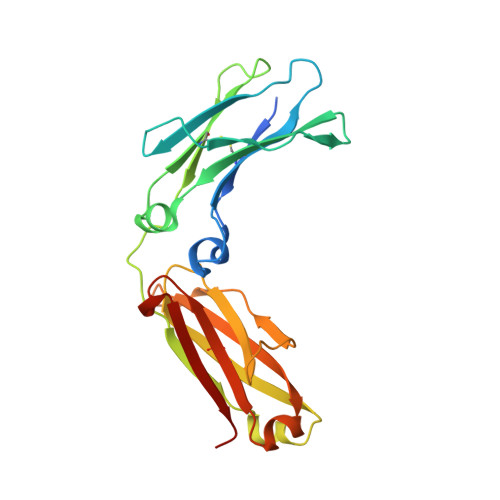
Crystal Structure of a Fc Fragment of Rhesus macaque (Macaca mulatta) IgG1.


Experimental Data Snapshot

  • Method: X-RAY DIFFRACTION
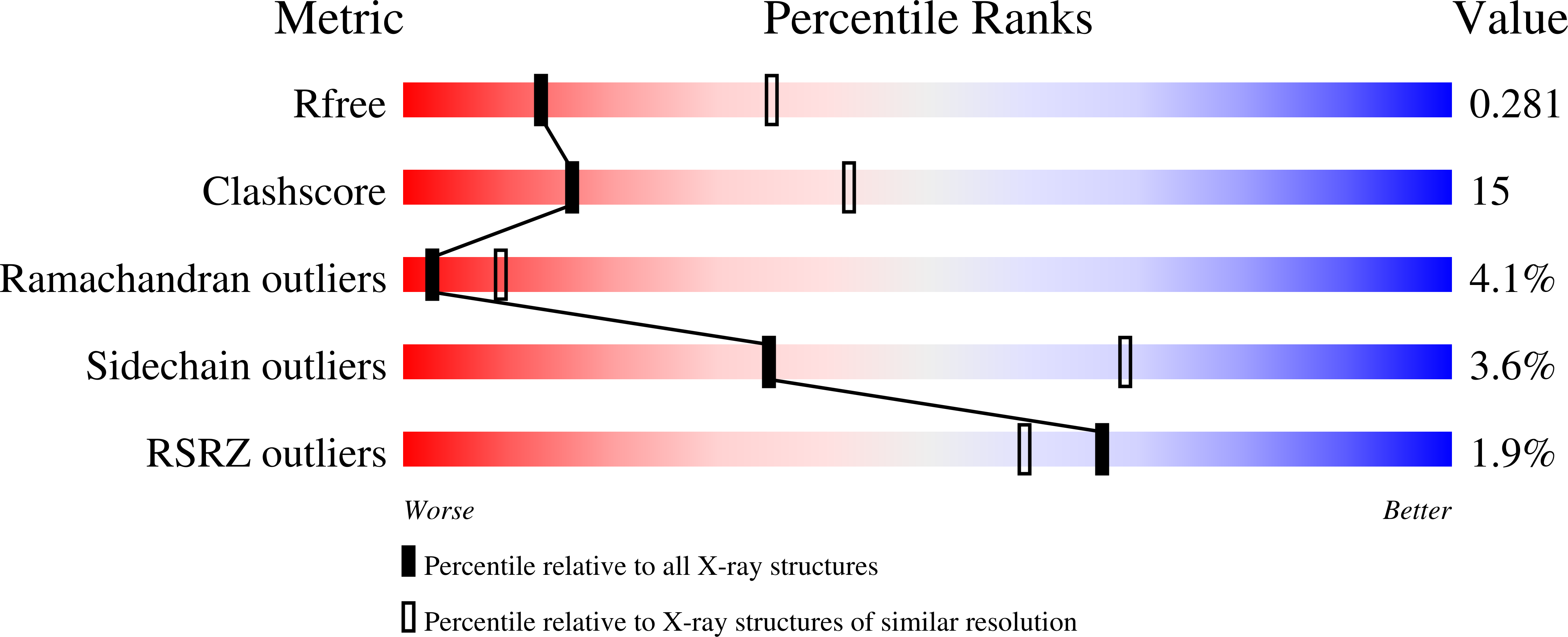
  • Resolution: 2.80 Å
  • R-Value Free: 0.272 
  • R-Value Work: 0.221 
  • R-Value Observed: 0.223 

Starting Model: experimental
View more details

wwPDB Validation   3D Report Full Report


This is version 2.2 of the entry. See complete history


Literature

From Rhesus macaque to human: structural evolutionary pathways for immunoglobulin G subclasses.

Tolbert, W.D.Subedi, G.P.Gohain, N.Lewis, G.K.Patel, K.R.Barb, A.W.Pazgier, M.

(2019) MAbs 11: 709-724

  • DOI: https://doi.org/10.1080/19420862.2019.1589852
  • Primary Citation of Related Structures:  
    6D4E, 6D4I, 6D4M, 6D4N

  • PubMed Abstract: 

    The Old World monkey, Rhesus macaque (Macaca mulatta, Mm), is frequently used as a primate model organism in the study of human disease and to test new vaccines/antibody treatments despite diverging before chimpanzees and orangutans. Mm and humans share 93% genome identity with substantial differences in the genes of the adaptive immune system that lead to different functional IgG subclass characteristics, Fcγ receptors expressed on innate immune cells, and biological interactions. These differences put limitations on Mm use as a primary animal model in the study of human disease and to test new vaccines/antibody treatments. Here, we comprehensively analyzed molecular properties of the Fc domain of the four IgG subclasses of Rhesus macaque to describe potential mechanisms for their interactions with effector cell Fc receptors. Our studies revealed less diversity in the overall structure among the Mm IgG Fc, with MmIgG1 Fc being the most structurally like human IgG3, although its C H 2 loops and N 297 glycan mobility are comparable to human IgG1. Furthermore, the Fcs of Mm IgG3 and 4 lack the structural properties typical for their human orthologues that determine IgG3's reduced interaction with the neonatal receptor and IgG4's ability for Fab-arm exchange and its weaker Fcγ receptor interactions. Taken together, our data indicate that MmIgG1-4 are less structurally divergent than the human IgGs, with only MmIgG1 matching the molecular properties of human IgG1 and 3, the most active IgGs in terms of Fcγ receptor binding and Fc-mediated functions. PDB accession numbers for deposited structures are 6D4E, 6D4I, 6D4M, and 6D4N for MmIgG1 Fc, MmIgG2 Fc, MmIgG3 Fc, and MmIgG4 Fc, respectively.


  • Organizational Affiliation

    a Division of Vaccine Research , Institute of Human Virology of University of Maryland School of Medicine , Baltimore , MD , USA.


Macromolecules
Find similar proteins by:  (by identity cutoff)  |  3D Structure
Entity ID: 1
MoleculeChains Sequence LengthOrganismDetailsImage
Fc Fragment of IgG1
A, B
224Macaca mulattaMutation(s): 0 
UniProt
Find proteins for F6RL33 (Macaca mulatta)
Explore F6RL33 
Go to UniProtKB:  F6RL33
Entity Groups  
Sequence Clusters30% Identity50% Identity70% Identity90% Identity95% Identity100% Identity
UniProt GroupF6RL33
Glycosylation
Glycosylation Sites: 1
Sequence Annotations
Expand
  • Reference Sequence
Oligosaccharides

Help

Entity ID: 2
MoleculeChains Length2D Diagram Glycosylation3D Interactions
2-acetamido-2-deoxy-beta-D-glucopyranose-(1-2)-alpha-D-mannopyranose-(1-3)-[2-acetamido-2-deoxy-beta-D-glucopyranose-(1-2)-alpha-D-mannopyranose-(1-6)]beta-D-mannopyranose-(1-4)-2-acetamido-2-deoxy-beta-D-glucopyranose-(1-4)-[alpha-L-fucopyranose-(1-6)]2-acetamido-2-deoxy-beta-D-glucopyranose
C, D
8N-Glycosylation
Glycosylation Resources
GlyTouCan:  G80858MF
GlyCosmos:  G80858MF
GlyGen:  G80858MF
Small Molecules
Ligands 1 Unique
IDChains Name / Formula / InChI Key2D Diagram3D Interactions
CL
Query on CL

Download Ideal Coordinates CCD File 
E [auth A]CHLORIDE ION
Cl
VEXZGXHMUGYJMC-UHFFFAOYSA-M
Experimental Data & Validation

Experimental Data

  • Method: X-RAY DIFFRACTION
  • Resolution: 2.80 Å
  • R-Value Free: 0.272 
  • R-Value Work: 0.221 
  • R-Value Observed: 0.223 
  • Space Group: P 1 21 1
Unit Cell:
Length ( Å )Angle ( ˚ )
a = 57.579α = 90
b = 64.525β = 104.55
c = 80.644γ = 90
Software Package:
Software NamePurpose
PHENIXrefinement
REFMACrefinement
HKL-2000data reduction
HKL-2000data scaling
PHASERphasing

Structure Validation

View Full Validation Report


Entry History & Funding Information

Deposition Data


Funding OrganizationLocationGrant Number
National Institutes of Health/National Institute Of Allergy and Infectious Diseases (NIH/NIAID)United StatesP01 AI20756

Revision History  (Full details and data files)

  • Version 1.0: 2019-05-01
    Type: Initial release
  • Version 1.1: 2019-06-05
    Changes: Data collection, Database references
  • Version 1.2: 2019-12-18
    Changes: Author supporting evidence, Data collection
  • Version 2.0: 2020-07-29
    Type: Remediation
    Reason: Carbohydrate remediation
    Changes: Atomic model, Data collection, Derived calculations, Structure summary
  • Version 2.1: 2023-10-04
    Changes: Data collection, Database references, Derived calculations, Refinement description, Structure summary
  • Version 2.2: 2024-10-16
    Changes: Structure summary