RCSB PDB - 6ZMA: Structure of the tRNA-Monooxygenase enzyme MiaE frozen under 140 bar of krypton using the soak and freeze methodology

 6ZMA

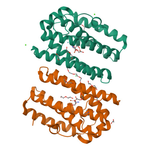
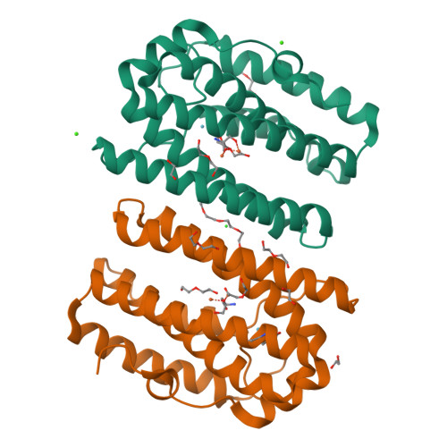
Structure of the tRNA-Monooxygenase enzyme MiaE frozen under 140 bar of krypton using the soak and freeze methodology


Experimental Data Snapshot

  • Method: X-RAY DIFFRACTION
  • Resolution: 2.15 Å
  • R-Value Free: 0.244 
  • R-Value Work: 0.180 
  • R-Value Observed: 0.183 

Starting Model: experimental
View more details

wwPDB Validation   3D Report Full Report


This is version 1.2 of the entry. See complete history


Literature

Structural, biochemical and functional analyses of tRNA-monooxygenase enzyme MiaE from Pseudomonas putida provide insights into tRNA/MiaE interaction.

Carpentier, P.Lepretre, C.Basset, C.Douki, T.Torelli, S.Duarte, V.Hamdane, D.Fontecave, M.Atta, M.

(2020) Nucleic Acids Res 48: 9918-9930

  • DOI: https://doi.org/10.1093/nar/gkaa667
  • Primary Citation of Related Structures:  
    6ZMA, 6ZMB, 6ZMC

  • PubMed Abstract: 

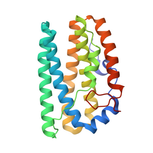
    MiaE (2-methylthio-N6-isopentenyl-adenosine37-tRNA monooxygenase) is a unique non-heme diiron enzyme that catalyzes the O2-dependent post-transcriptional allylic hydroxylation of a hypermodified nucleotide 2-methylthio-N6-isopentenyl-adenosine (ms2i6A37) at position 37 of selected tRNA molecules to produce 2-methylthio-N6-4-hydroxyisopentenyl-adenosine (ms2io6A37). Here, we report the in vivo activity, biochemical, spectroscopic characterization and X-ray crystal structure of MiaE from Pseudomonas putida. The investigation demonstrates that the putative pp-2188 gene encodes a MiaE enzyme. The structure shows that Pp-MiaE consists of a catalytic diiron(III) domain with a four alpha-helix bundle fold. A docking model of Pp-MiaE in complex with tRNA, combined with site directed mutagenesis and in vivo activity shed light on the importance of an additional linker region for substrate tRNA recognition. Finally, krypton-pressurized Pp-MiaE experiments, revealed the presence of defined O2 site along a conserved hydrophobic tunnel leading to the diiron active center.


  • Organizational Affiliation

    Univ. Grenoble Alpes, CEA, CNRS, CBM-UMR 5249, 17 avenue des martyrs, Grenoble, France.


Macromolecules
Find similar proteins by:  (by identity cutoff)  |  3D Structure
Entity ID: 1
MoleculeChains Sequence LengthOrganismDetailsImage
tRNA hydroxylaseA [auth B],
B [auth C]
205Pseudomonas putidaMutation(s): 0 
Gene Names: AYO08_21560CBL13_00280CBP06_18695
UniProt
Find proteins for A0A179QS89 (Pseudomonas putida)
Explore A0A179QS89 
Go to UniProtKB:  A0A179QS89
Entity Groups  
Sequence Clusters30% Identity50% Identity70% Identity90% Identity95% Identity100% Identity
UniProt GroupA0A179QS89
Sequence Annotations
Expand
  • Reference Sequence
Small Molecules
Ligands 8 Unique
IDChains Name / Formula / InChI Key2D Diagram3D Interactions
PG4
Query on PG4

Download Ideal Coordinates CCD File 
L [auth B]TETRAETHYLENE GLYCOL
C8 H18 O5
UWHCKJMYHZGTIT-UHFFFAOYSA-N
TRS
Query on TRS

Download Ideal Coordinates CCD File 
H [auth B],
R [auth C]
2-AMINO-2-HYDROXYMETHYL-PROPANE-1,3-DIOL
C4 H12 N O3
LENZDBCJOHFCAS-UHFFFAOYSA-O
PEG
Query on PEG

Download Ideal Coordinates CCD File 
I [auth B]
J [auth B]
K [auth B]
S [auth C]
T [auth C]
DI(HYDROXYETHYL)ETHER
C4 H10 O3
MTHSVFCYNBDYFN-UHFFFAOYSA-N
KR (Subject of Investigation/LOI)
Query on KR

Download Ideal Coordinates CCD File 
M [auth B]
N [auth B]
W [auth C]
X [auth C]
Y [auth C]
KRYPTON
Kr
DNNSSWSSYDEUBZ-UHFFFAOYSA-N
EDO
Query on EDO

Download Ideal Coordinates CCD File 
AA [auth C],
BA [auth C],
O [auth B]
1,2-ETHANEDIOL
C2 H6 O2
LYCAIKOWRPUZTN-UHFFFAOYSA-N
FE (Subject of Investigation/LOI)
Query on FE

Download Ideal Coordinates CCD File 
C [auth B],
D [auth B],
P [auth C],
Q [auth C]
FE (III) ION
Fe
VTLYFUHAOXGGBS-UHFFFAOYSA-N
CA
Query on CA

Download Ideal Coordinates CCD File 
F [auth B],
G [auth B]
CALCIUM ION
Ca
BHPQYMZQTOCNFJ-UHFFFAOYSA-N
CL
Query on CL

Download Ideal Coordinates CCD File 
E [auth B]CHLORIDE ION
Cl
VEXZGXHMUGYJMC-UHFFFAOYSA-M
Experimental Data & Validation

Experimental Data

  • Method: X-RAY DIFFRACTION
  • Resolution: 2.15 Å
  • R-Value Free: 0.244 
  • R-Value Work: 0.180 
  • R-Value Observed: 0.183 
  • Space Group: C 1 2 1
Unit Cell:
Length ( Å )Angle ( ˚ )
a = 117.622α = 90
b = 52.3β = 90.74
c = 79.407γ = 90
Software Package:
Software NamePurpose
REFMACrefinement
PDB_EXTRACTdata extraction
XDSdata reduction
XSCALEdata scaling
PHASERphasing

Structure Validation

View Full Validation Report


Entry History & Funding Information

Deposition Data


Funding OrganizationLocationGrant Number
French National Research AgencyFranceANR-15-CE21-0020

Revision History  (Full details and data files)

  • Version 1.0: 2020-09-02
    Type: Initial release
  • Version 1.1: 2020-10-07
    Changes: Database references
  • Version 1.2: 2024-01-31
    Changes: Advisory, Data collection, Database references, Refinement description