Assisted assembly of bacteriophage T7 core components for genome translocation across the bacterial envelope.
Perez-Ruiz, M., Pulido-Cid, M., Luque-Ortega, J.R., Valpuesta, J.M., Cuervo, A., Carrascosa, J.L.(2021) Proc Natl Acad Sci U S A 118
- PubMed: 34417311
- DOI: https://doi.org/10.1073/pnas.2026719118
- Primary Citation of Related Structures:
6YSZ, 6YT5 - PubMed Abstract:
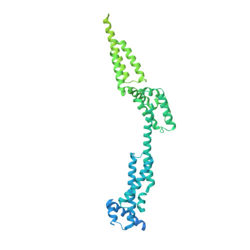
In most bacteriophages, genome transport across bacterial envelopes is carried out by the tail machinery. In viruses of the Podoviridae family, in which the tail is not long enough to traverse the bacterial wall, it has been postulated that viral core proteins assembled inside the viral head are translocated and reassembled into a tube within the periplasm that extends the tail channel. Bacteriophage T7 infects Escherichia coli , and despite extensive studies, the precise mechanism by which its genome is translocated remains unknown. Using cryo-electron microscopy, we have resolved the structure of two different assemblies of the T7 DNA translocation complex composed of the core proteins gp15 and gp16. Gp15 alone forms a partially folded hexamer, which is further assembled upon interaction with gp16 into a tubular structure, forming a channel that could allow DNA passage. The structure of the gp15-gp16 complex also shows the location within gp16 of a canonical transglycosylase motif involved in the degradation of the bacterial peptidoglycan layer. This complex docks well in the tail extension structure found in the periplasm of T7-infected bacteria and matches the sixfold symmetry of the phage tail. In such cases, gp15 and gp16 that are initially present in the T7 capsid eightfold-symmetric core would change their oligomeric state upon reassembly in the periplasm. Altogether, these results allow us to propose a model for the assembly of the core translocation complex in the periplasm, which furthers understanding of the molecular mechanism involved in the release of T7 viral DNA into the bacterial cytoplasm.
Organizational Affiliation:
Centro Nacional de Biotecnología, Madrid 28049, Spain.


