6V2D

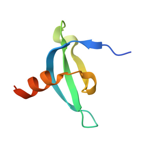
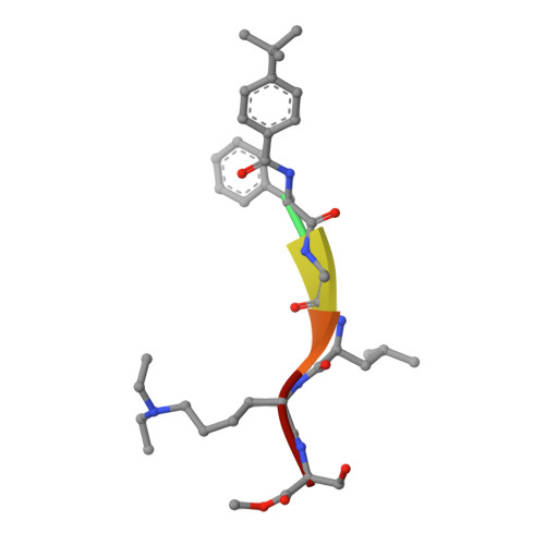
Crystal Structure of chromodomain of CDYL2 in complex with inhibitor UNC3866


Experimental Data Snapshot

  • Method: X-RAY DIFFRACTION
  • Resolution: 2.10 Å
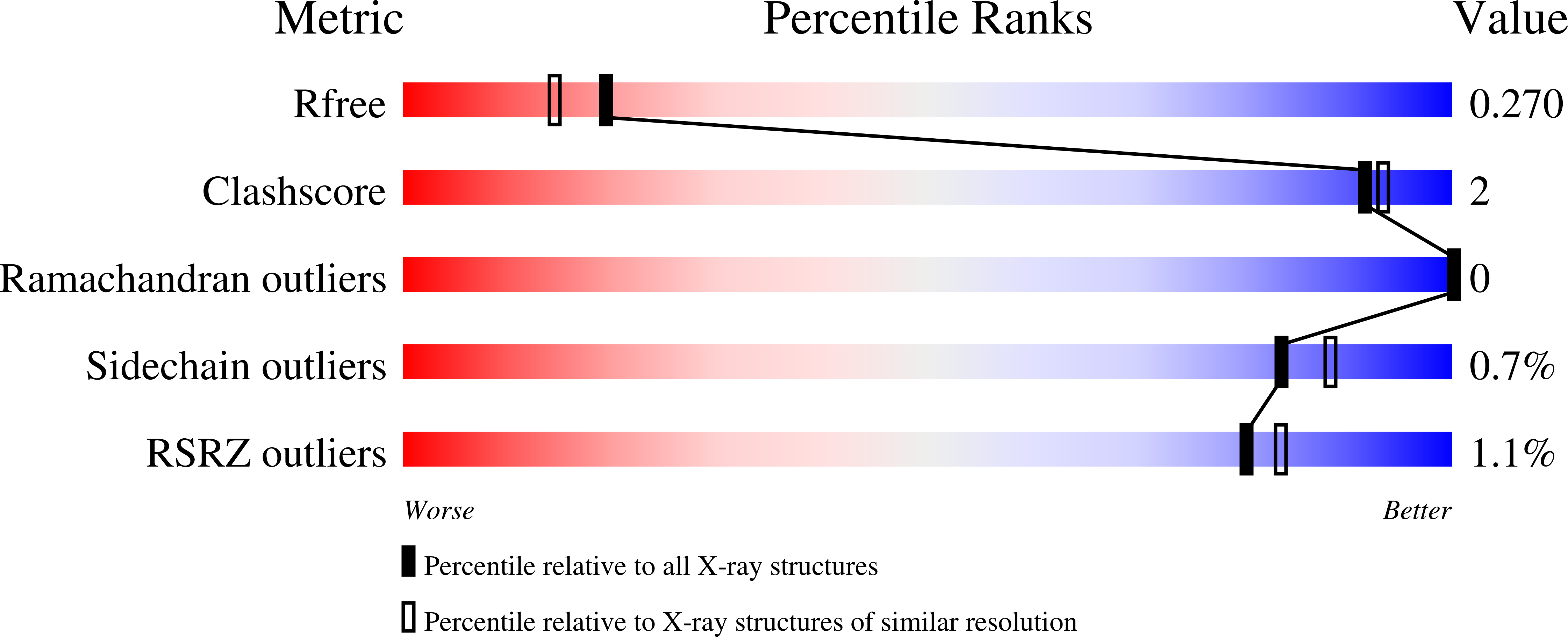
  • R-Value Free: 0.268 
  • R-Value Work: 0.212 
  • R-Value Observed: 0.215 

Starting Models: experimental
View more details

wwPDB Validation   3D Report Full Report


This is version 1.3 of the entry. See complete history


Literature

Structural Basis for the Binding Selectivity of Human CDY Chromodomains.

Dong, C.Liu, Y.Lyu, T.J.Beldar, S.Lamb, K.N.Tempel, W.Li, Y.Li, Z.James, L.I.Qin, S.Wang, Y.Min, J.

(2020) Cell Chem Biol 27: 827-838.e7

  • DOI: https://doi.org/10.1016/j.chembiol.2020.05.007
  • Primary Citation of Related Structures:  
    2MJ8, 6V2D, 6V2H, 6V2R, 6V2S, 6V3N, 6V41, 6V8W

  • PubMed Abstract: 

    The CDY (chromodomain on the Y) proteins play an essential role in normal spermatogenesis and brain development. Dysregulation of their expression has been linked to male infertility and various neurological diseases. Like the chromodomains of HP1 and Polycomb, the CDY chromodomains also recognize the lysine-methylated ARKS motif embedded in histone and non-histone proteins. Interestingly, the CDY chromodomains exhibit different binding preferences for the lysine-methylated ARKS motif in different sequence contexts. Here, we present the structural basis for selective binding of CDY1 to H3K9me3 and preferential binding of CDYL2 to H3tK27me3 over H3K27me3. In addition, we use a CDYL1/2-selective compound, UNC4850, to gain further insight into the molecular mechanisms underlying CDYL2 binding specificity. Our work also provides critical implications that CDYL1b's role in the regulation of neural development is dependent on its recognition of the lysine-methylated ARKS motif.


  • Organizational Affiliation

    Department of Biochemistry and Molecular Biology, School of Basic Medical Sciences, Tianjin Medical University, Tianjin 300070, China.


Macromolecules
Find similar proteins by:  (by identity cutoff)  |  3D Structure
Entity ID: 1
MoleculeChains Sequence LengthOrganismDetailsImage
Chromodomain Y-like protein 264Homo sapiensMutation(s): 0 
Gene Names: CDYL2
UniProt & NIH Common Fund Data Resources
Find proteins for Q8N8U2 (Homo sapiens)
Explore Q8N8U2 
Go to UniProtKB:  Q8N8U2
PHAROS:  Q8N8U2
GTEx:  ENSG00000166446 
Entity Groups  
Sequence Clusters30% Identity50% Identity70% Identity90% Identity95% Identity100% Identity
UniProt GroupQ8N8U2
Sequence Annotations
Expand
  • Reference Sequence

Find similar proteins by:  Sequence   |   3D Structure  

Entity ID: 2
MoleculeChains Sequence LengthOrganismDetailsImage
UNC38666synthetic constructMutation(s): 0 
Sequence Annotations
Expand
  • Reference Sequence
Small Molecules
Ligands 1 Unique
IDChains Name / Formula / InChI Key2D Diagram3D Interactions
UNX
Query on UNX

Download Ideal Coordinates CCD File 
AA [auth G]
BA [auth G]
CA [auth G]
DA [auth G]
EA [auth G]
AA [auth G],
BA [auth G],
CA [auth G],
DA [auth G],
EA [auth G],
FA [auth G],
GA [auth I],
HA [auth I],
IA [auth I],
JA [auth I],
KA [auth I],
LA [auth K],
M [auth A],
MA [auth K],
N [auth A],
NA [auth J],
O [auth A],
P [auth A],
Q [auth A],
R [auth A],
S [auth A],
T [auth A],
U [auth C],
V [auth C],
W [auth C],
X [auth C],
Y [auth C],
Z [auth E]
UNKNOWN ATOM OR ION
X
Modified Residues  2 Unique
IDChains TypeFormula2D DiagramParent
5R5
Query on 5R5
G [auth J]
H [auth L]
I [auth B]
J [auth D]
K [auth F]
G [auth J],
H [auth L],
I [auth B],
J [auth D],
K [auth F],
L [auth H]
L-PEPTIDE LINKINGC4 H9 N O3SER
ELY
Query on ELY
G [auth J]
H [auth L]
I [auth B]
J [auth D]
K [auth F]
G [auth J],
H [auth L],
I [auth B],
J [auth D],
K [auth F],
L [auth H]
L-PEPTIDE LINKINGC10 H22 N2 O2LYS
Experimental Data & Validation

Experimental Data

Unit Cell:
Length ( Å )Angle ( ˚ )
a = 45.979α = 90
b = 83.835β = 90
c = 115.288γ = 90
Software Package:
Software NamePurpose
PHENIXrefinement
Aimlessdata scaling
PDB_EXTRACTdata extraction
XDSdata reduction
PHASERphasing

Structure Validation

View Full Validation Report


Entry History 

Deposition Data

Revision History  (Full details and data files)

  • Version 1.0: 2019-12-25
    Type: Initial release
  • Version 1.1: 2020-06-17
    Changes: Database references
  • Version 1.2: 2020-07-29
    Changes: Database references
  • Version 1.3: 2023-10-11
    Changes: Data collection, Database references, Refinement description