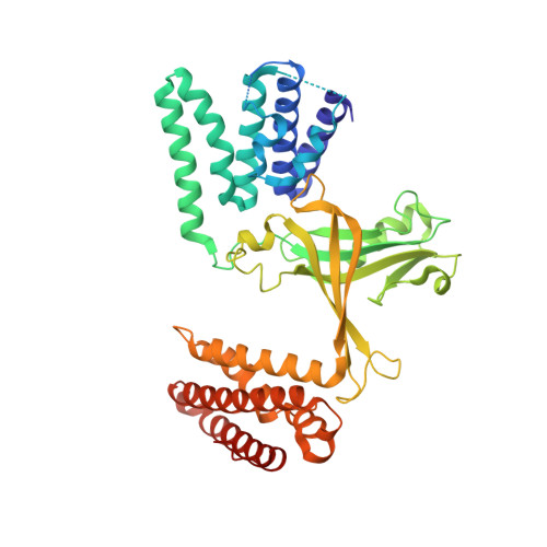
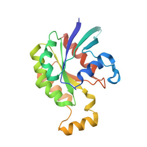
Structural basis for CDC42 and RAC activation by the dual specificity GEF DOCK10
Fan, D., Yang, J., Cronin, N., Barford, D.(2022) bioRxiv
Experimental Data Snapshot
Starting Model: experimental
View more details
(2022) bioRxiv
Entity ID: 1 | |||||
|---|---|---|---|---|---|
| Molecule | Chains | Sequence Length | Organism | Details | Image |
| Dedicator of cytokinesis protein 10 | A [auth B], D [auth A] | 458 | Homo sapiens | Mutation(s): 0 Gene Names: DOCK10, KIAA0694, ZIZ3 |  |
UniProt & NIH Common Fund Data Resources | |||||
Find proteins for Q96BY6 (Homo sapiens) Explore Q96BY6 Go to UniProtKB: Q96BY6 | |||||
PHAROS: Q96BY6 GTEx: ENSG00000135905 | |||||
Entity Groups | |||||
| Sequence Clusters | 30% Identity50% Identity70% Identity90% Identity95% Identity100% Identity | ||||
| UniProt Group | Q96BY6 | ||||
Sequence AnnotationsExpand | |||||
| |||||
Entity ID: 2 | |||||
|---|---|---|---|---|---|
| Molecule | Chains | Sequence Length | Organism | Details | Image |
| Cell division control protein 42 homolog | B [auth C], C [auth D] | 188 | Homo sapiens | Mutation(s): 0 Gene Names: CDC42 EC: 3.6.5.2 |  |
UniProt & NIH Common Fund Data Resources | |||||
Find proteins for P60953 (Homo sapiens) Explore P60953 Go to UniProtKB: P60953 | |||||
PHAROS: P60953 GTEx: ENSG00000070831 | |||||
Entity Groups | |||||
| Sequence Clusters | 30% Identity50% Identity70% Identity90% Identity95% Identity100% Identity | ||||
| UniProt Group | P60953 | ||||
Sequence AnnotationsExpand | |||||
| |||||
| Ligands 2 Unique | |||||
|---|---|---|---|---|---|
| ID | Chains | Name / Formula / InChI Key | 2D Diagram | 3D Interactions | |
| GDP (Subject of Investigation/LOI) Query on GDP | E [auth C], F [auth D] | GUANOSINE-5'-DIPHOSPHATE C10 H15 N5 O11 P2 QGWNDRXFNXRZMB-UUOKFMHZSA-N |  | ||
| GOL (Subject of Investigation/LOI) Query on GOL | G [auth A] | GLYCEROL C3 H8 O3 PEDCQBHIVMGVHV-UHFFFAOYSA-N |  | ||
| Length ( Å ) | Angle ( ˚ ) |
|---|---|
| a = 97.17 | α = 90 |
| b = 97.17 | β = 90 |
| c = 312 | γ = 90 |
| Software Name | Purpose |
|---|---|
| PHENIX | refinement |
| PHENIX | refinement |
| MOSFLM | data reduction |
| SCALA | data scaling |
| PHASER | phasing |
| Funding Organization | Location | Grant Number |
|---|---|---|
| Medical Research Council (MRC, United Kingdom) | United Kingdom | MC_UP_1201/6 |
| Cancer Research UK | United Kingdom | C576/A14109 |