6P1Y

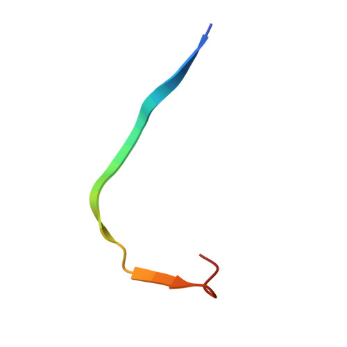
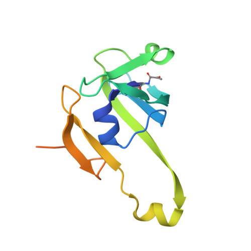
Crystal structure of Mtb aspartate decarboxylase mutant M117I


Experimental Data Snapshot

  • Method: X-RAY DIFFRACTION
  • Resolution: 2.33 Å
  • R-Value Free: 0.204 
  • R-Value Work: 0.174 
  • R-Value Observed: 0.176 

Starting Model: experimental
View more details

wwPDB Validation   3D Report Full Report


This is version 2.1 of the entry. See complete history


Literature

The molecular basis of pyrazinamide activity on Mycobacterium tuberculosis PanD.

Sun, Q.Li, X.Perez, L.M.Shi, W.Zhang, Y.Sacchettini, J.C.

(2020) Nat Commun 11: 339-339

  • DOI: https://doi.org/10.1038/s41467-019-14238-3
  • Primary Citation of Related Structures:  
    6OYY, 6OZ8, 6P02, 6P1Y

  • PubMed Abstract: 

    Pyrazinamide has been a mainstay in the multidrug regimens used to treat tuberculosis. It is active against the persistent, non-replicating mycobacteria responsible for the protracted therapy required to cure tuberculosis. Pyrazinamide is a pro-drug that is converted into pyrazinoic acid (POA) by pyrazinamidase, however, the exact target of the drug has been difficult to determine. Here we show the enzyme PanD binds POA in its active site in a manner consistent with competitive inhibition. The active site is not directly accessible to the inhibitor, suggesting the protein must undergo a conformational change to bind the inhibitor. This is consistent with the slow binding kinetics we determined for POA. Drug-resistant mutations cluster near loops that lay on top of the active site. These resistant mutants show reduced affinity and residence time of POA consistent with a model where resistance occurs by destabilizing the closed conformation of the active site.


  • Organizational Affiliation

    Department of Biochemistry and Biophysics, Texas A&M University, College Station, TX, USA.


Macromolecules

Find similar proteins by:  Sequence   |   3D Structure  

Entity ID: 1
MoleculeChains Sequence LengthOrganismDetailsImage
Aspartate 1-decarboxylase beta chain24Mycobacterium tuberculosis H37RvMutation(s): 0 
Gene Names: panDRv3601cMTCY07H7B.21
EC: 4.1.1.11
UniProt
Find proteins for P9WIL3 (Mycobacterium tuberculosis (strain ATCC 25618 / H37Rv))
Explore P9WIL3 
Go to UniProtKB:  P9WIL3
Entity Groups  
Sequence Clusters30% Identity50% Identity70% Identity90% Identity95% Identity100% Identity
UniProt GroupP9WIL3
Sequence Annotations
Expand
  • Reference Sequence
Find similar proteins by:  (by identity cutoff)  |  3D Structure
Entity ID: 2
MoleculeChains Sequence LengthOrganismDetailsImage
Aspartate 1-decarboxylase alpha chain123Mycobacterium tuberculosis H37RvMutation(s): 2 
Gene Names: panDRv3601cMTCY07H7B.21
EC: 4.1.1.11
UniProt
Find proteins for P9WIL3 (Mycobacterium tuberculosis (strain ATCC 25618 / H37Rv))
Explore P9WIL3 
Go to UniProtKB:  P9WIL3
Entity Groups  
Sequence Clusters30% Identity50% Identity70% Identity90% Identity95% Identity100% Identity
UniProt GroupP9WIL3
Sequence Annotations
Expand
  • Reference Sequence
Experimental Data & Validation

Experimental Data

  • Method: X-RAY DIFFRACTION
  • Resolution: 2.33 Å
  • R-Value Free: 0.204 
  • R-Value Work: 0.174 
  • R-Value Observed: 0.176 
  • Space Group: I 4 2 2
Unit Cell:
Length ( Å )Angle ( ˚ )
a = 73.011α = 90
b = 73.011β = 90
c = 109.53γ = 90
Software Package:
Software NamePurpose
PHENIXrefinement
HKL-3000data reduction
HKL-3000data scaling
PHENIXphasing

Structure Validation

View Full Validation Report


Entry History & Funding Information

Deposition Data


Funding OrganizationLocationGrant Number
National Institutes of Health/National Institute Of Allergy and Infectious Diseases (NIH/NIAID)United StatesP01AI095208
Welch FoundationUnited StatesA-0015

Revision History  (Full details and data files)

  • Version 1.0: 2020-02-05
    Type: Initial release
  • Version 1.1: 2023-10-11
    Changes: Data collection, Database references, Refinement description
  • Version 2.0: 2023-11-15
    Changes: Atomic model, Data collection, Derived calculations
  • Version 2.1: 2024-10-23
    Changes: Structure summary