6OB2

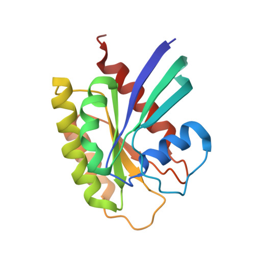
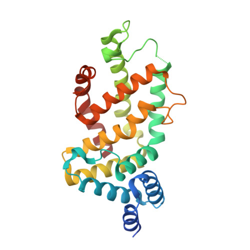
Crystal structure of wild-type KRAS (GMPPNP-bound) in complex with GAP-related domain (GRD) of neurofibromin (NF1)


Experimental Data Snapshot

  • Method: X-RAY DIFFRACTION
  • Resolution: 2.85 Å
  • R-Value Free: 0.247 
  • R-Value Work: 0.199 
  • R-Value Observed: 0.202 

Starting Model: experimental
View more details

wwPDB Validation   3D Report Full Report


Ligand Structure Quality Assessment 


This is version 1.3 of the entry. See complete history


Literature

KRAS G13D sensitivity to neurofibromin-mediated GTP hydrolysis.

Rabara, D.Tran, T.H.Dharmaiah, S.Stephens, R.M.McCormick, F.Simanshu, D.K.Holderfield, M.

(2019) Proc Natl Acad Sci U S A 116: 22122-22131

  • DOI: https://doi.org/10.1073/pnas.1908353116
  • Primary Citation of Related Structures:  
    6OB2, 6OB3

  • PubMed Abstract: 

    KRAS mutations occur in ∼35% of colorectal cancers and promote tumor growth by constitutively activating the mitogen-activated protein kinase (MAPK) pathway. KRAS mutations at codons 12, 13, or 61 are thought to prevent GAP protein-stimulated GTP hydrolysis and render KRAS -mutated colorectal cancers unresponsive to epidermal growth factor receptor (EGFR) inhibitors. We report here that KRAS G13-mutated cancer cells are frequently comutated with NF1 GAP but NF1 is rarely mutated in cancers with KRAS codon 12 or 61 mutations. Neurofibromin protein (encoded by the NF1 gene) hydrolyzes GTP directly in complex with KRAS G13D, and KRAS G13D-mutated cells can respond to EGFR inhibitors in a neurofibromin-dependent manner. Structures of the wild type and G13D mutant of KRAS in complex with neurofibromin (RasGAP domain) provide the structural basis for neurofibromin-mediated GTP hydrolysis. These results reveal that KRAS G13D is responsive to neurofibromin-stimulated hydrolysis and suggest that a subset of KRAS G13-mutated colorectal cancers that are neurofibromin-competent may respond to EGFR therapies.


  • Organizational Affiliation

    NCI RAS Initiative, Cancer Research Technology Program, Frederick National Laboratory for Cancer Research, Frederick, MD 21701.


Macromolecules
Find similar proteins by:  (by identity cutoff)  |  3D Structure
Entity ID: 1
MoleculeChains Sequence LengthOrganismDetailsImage
GTPase KRas
A, C
170Homo sapiensMutation(s): 0 
Gene Names: KRASKRAS2RASK2
EC: 3.6.5.2
UniProt & NIH Common Fund Data Resources
Find proteins for P01116 (Homo sapiens)
Explore P01116 
Go to UniProtKB:  P01116
PHAROS:  P01116
GTEx:  ENSG00000133703 
Entity Groups  
Sequence Clusters30% Identity50% Identity70% Identity90% Identity95% Identity100% Identity
UniProt GroupP01116
Sequence Annotations
Expand
  • Reference Sequence
Find similar proteins by:  (by identity cutoff)  |  3D Structure
Entity ID: 2
MoleculeChains Sequence LengthOrganismDetailsImage
Neurofibromin
B, D
256Homo sapiensMutation(s): 0 
Gene Names: NF1
UniProt & NIH Common Fund Data Resources
Find proteins for P21359 (Homo sapiens)
Explore P21359 
Go to UniProtKB:  P21359
PHAROS:  P21359
GTEx:  ENSG00000196712 
Entity Groups  
Sequence Clusters30% Identity50% Identity70% Identity90% Identity95% Identity100% Identity
UniProt GroupP21359
Sequence Annotations
Expand
  • Reference Sequence
Small Molecules
Ligands 5 Unique
IDChains Name / Formula / InChI Key2D Diagram3D Interactions
GNP (Subject of Investigation/LOI)
Query on GNP

Download Ideal Coordinates CCD File 
F [auth A],
K [auth C]
PHOSPHOAMINOPHOSPHONIC ACID-GUANYLATE ESTER
C10 H17 N6 O13 P3
UQABYHGXWYXDTK-UUOKFMHZSA-N
GOL
Query on GOL

Download Ideal Coordinates CCD File 
E [auth A],
H [auth B]
GLYCEROL
C3 H8 O3
PEDCQBHIVMGVHV-UHFFFAOYSA-N
IMD
Query on IMD

Download Ideal Coordinates CCD File 
I [auth B]IMIDAZOLE
C3 H5 N2
RAXXELZNTBOGNW-UHFFFAOYSA-O
CL
Query on CL

Download Ideal Coordinates CCD File 
J [auth B]CHLORIDE ION
Cl
VEXZGXHMUGYJMC-UHFFFAOYSA-M
MG (Subject of Investigation/LOI)
Query on MG

Download Ideal Coordinates CCD File 
G [auth A],
L [auth C]
MAGNESIUM ION
Mg
JLVVSXFLKOJNIY-UHFFFAOYSA-N
Experimental Data & Validation

Experimental Data

  • Method: X-RAY DIFFRACTION
  • Resolution: 2.85 Å
  • R-Value Free: 0.247 
  • R-Value Work: 0.199 
  • R-Value Observed: 0.202 
  • Space Group: P 21 21 21
Unit Cell:
Length ( Å )Angle ( ˚ )
a = 83.728α = 90
b = 99.736β = 90
c = 155.514γ = 90
Software Package:
Software NamePurpose
XDSdata reduction
XSCALEdata scaling
PHASERphasing
PHENIXrefinement
PDB_EXTRACTdata extraction

Structure Validation

View Full Validation Report


Ligand Structure Quality Assessment 


Entry History 

Deposition Data

Revision History  (Full details and data files)

  • Version 1.0: 2019-10-16
    Type: Initial release
  • Version 1.1: 2019-10-30
    Changes: Data collection, Database references
  • Version 1.2: 2019-11-13
    Changes: Data collection, Database references
  • Version 1.3: 2023-10-11
    Changes: Data collection, Database references, Derived calculations, Refinement description