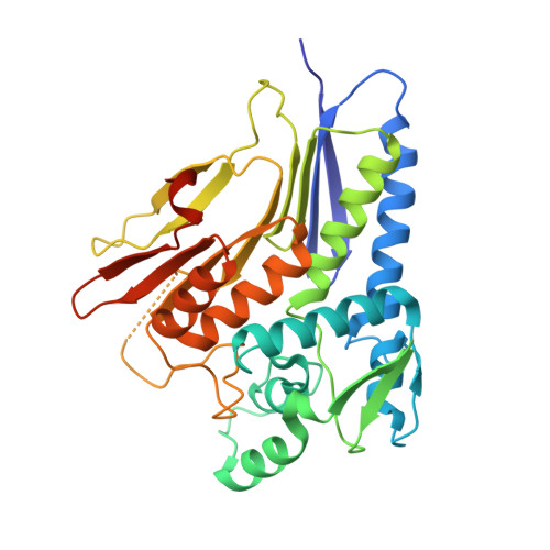
Conserved residue His-257 ofVibrio choleraeflavin transferase ApbE plays a critical role in substrate binding and catalysis.
Fang, X., Osipiuk, J., Chakravarthy, S., Yuan, M., Menzer, W.M., Nissen, D., Liang, P., Raba, D.A., Tuz, K., Howard, A.J., Joachimiak, A., Minh, D.D.L., Juarez, O.(2019) J Biol Chem 294: 13800-13810
- PubMed: 31350338
- DOI: https://doi.org/10.1074/jbc.RA119.008261
- Primary Citation of Related Structures:
6NXI, 6NXJ - PubMed Abstract:
The flavin transferase ApbE plays essential roles in bacterial physiology, covalently incorporating FMN cofactors into numerous respiratory enzymes that use the integrated cofactors as electron carriers. In this work we performed a detailed kinetic and structural characterization of Vibrio cholerae WT ApbE and mutants of the conserved residue His-257, to understand its role in substrate binding and in the catalytic mechanism of this family. Bi-substrate kinetic experiments revealed that ApbE follows a random Bi Bi sequential kinetic mechanism, in which a ternary complex is formed, indicating that both substrates must be bound to the enzyme for the reaction to proceed. Steady-state kinetic analyses show that the turnover rates of His-257 mutants are significantly smaller than those of WT ApbE, and have increased K m values for both substrates, indicating that the His-257 residue plays important roles in catalysis and in enzyme-substrate complex formation. Analyses of the pH dependence of ApbE activity indicate that the p K a of the catalytic residue (p K ES1 ) increases by 2 pH units in the His-257 mutants, suggesting that this residue plays a role in substrate deprotonation. The crystal structures of WT ApbE and an H257G mutant were determined at 1.61 and 1.92 Å resolutions, revealing that His-257 is located in the catalytic site and that the substitution does not produce major conformational changes. We propose a reaction mechanism in which His-257 acts as a general base that deprotonates the acceptor residue, which subsequently performs a nucleophilic attack on FAD for flavin transfer.
Organizational Affiliation:
Department of Biological Sciences, Illinois Institute of Technology, Chicago, Illinois 60616.


