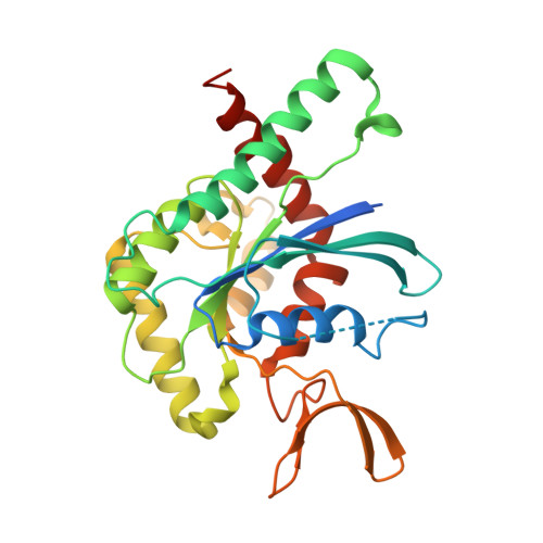
A complete compendium of crystal structures for the human SEPT3 subgroup reveals functional plasticity at a specific septin interface.
Castro, D.K.S.D.V., da Silva, S.M.O., Pereira, H.D., Macedo, J.N.A., Leonardo, D.A., Valadares, N.F., Kumagai, P.S., Brandao-Neto, J., Araujo, A.P.U., Garratt, R.C.(2020) IUCrJ 7: 462-479
- PubMed: 32431830
- DOI: https://doi.org/10.1107/S2052252520002973
- Primary Citation of Related Structures:
6MQ9, 6MQB, 6MQK - PubMed Abstract:
Human septins 3, 9 and 12 are the only members of a specific subgroup of septins that display several unusual features, including the absence of a C-terminal coiled coil. This particular subgroup (the SEPT3 septins) are present in rod-like octameric protofilaments but are lacking in similar hexameric assemblies, which only contain representatives of the three remaining subgroups. Both hexamers and octamers can self-assemble into mixed filaments by end-to-end association, implying that the SEPT3 septins may facilitate polymerization but not necessarily function. These filaments frequently associate into higher order complexes which associate with biological membranes, triggering a wide range of cellular events. In the present work, a complete compendium of crystal structures for the GTP-binding domains of all of the SEPT3 subgroup members when bound to either GDP or to a GTP analogue is provided. The structures reveal a unique degree of plasticity at one of the filamentous interfaces (dubbed NC). Specifically, structures of the GDP and GTPγS complexes of SEPT9 reveal a squeezing mechanism at the NC interface which would expel a polybasic region from its binding site and render it free to interact with negatively charged membranes. On the other hand, a polyacidic region associated with helix α5', the orientation of which is particular to this subgroup, provides a safe haven for the polybasic region when retracted within the interface. Together, these results suggest a mechanism which couples GTP binding and hydrolysis to membrane association and implies a unique role for the SEPT3 subgroup in this process. These observations can be accounted for by constellations of specific amino-acid residues that are found only in this subgroup and by the absence of the C-terminal coiled coil. Such conclusions can only be reached owing to the completeness of the structural studies presented here.
Organizational Affiliation:
Instituto de Física de São Carlos, Universidade de São Paulo, Avenida Joao Dagnone 1100, São Carlos-SP 13563-723, Brazil.


