6KMP

100K X-ray structure of HIV-1 protease triple mutant (V32I,I47V,V82I) with tetrahedral intermediate mimic KVS-1


Experimental Data Snapshot

  • Method: X-RAY DIFFRACTION
  • Resolution: 1.31 Å
  • R-Value Free: 0.188 
  • R-Value Work: 0.156 
  • R-Value Observed: 0.158 

Starting Model: experimental
View more details

wwPDB Validation   3D Report Full Report


Ligand Structure Quality Assessment 


This is version 1.1 of the entry. See complete history


Literature

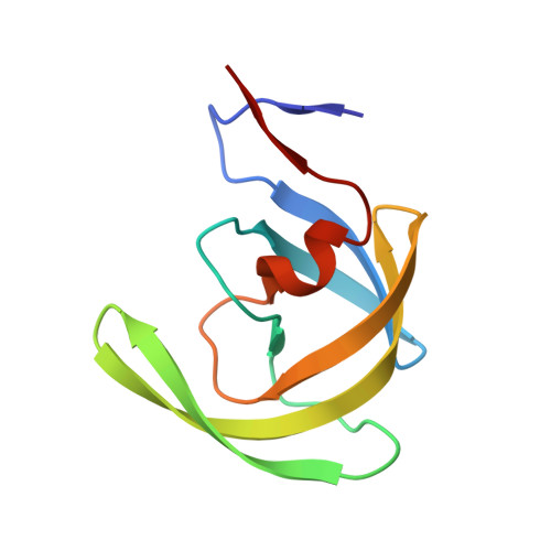
Visualizing Tetrahedral Oxyanion Bound in HIV-1 Protease Using Neutrons: Implications for the Catalytic Mechanism and Drug Design.

Kumar, M.Mandal, K.Blakeley, M.P.Wymore, T.Kent, S.B.H.Louis, J.M.Das, A.Kovalevsky, A.

(2020) ACS Omega 5: 11605-11617

  • DOI: https://doi.org/10.1021/acsomega.0c00835
  • Primary Citation of Related Structures:  
    6KMP, 6PTP, 6PU8

  • PubMed Abstract: 

    HIV-1 protease is indispensable for virus propagation and an important therapeutic target for antiviral inhibitors to treat AIDS. As such inhibitors are transition-state mimics, a detailed understanding of the enzyme mechanism is crucial for the development of better anti-HIV drugs. Here, we used room-temperature joint X-ray/neutron crystallography to directly visualize hydrogen atoms and map hydrogen bonding interactions in a protease complex with peptidomimetic inhibitor KVS-1 containing a reactive nonhydrolyzable ketomethylene isostere, which, upon reacting with the catalytic water molecule, is converted into a tetrahedral intermediate state, KVS-1 TI . We unambiguously determined that the resulting tetrahedral intermediate is an oxyanion, rather than the gem -diol, and both catalytic aspartic acid residues are protonated. The oxyanion tetrahedral intermediate appears to be unstable, even though the negative charge on the oxyanion is delocalized through a strong n → π* hyperconjugative interaction into the nearby peptidic carbonyl group of the inhibitor. To better understand the influence of the ketomethylene isostere as a protease inhibitor, we have also examined the protease structure and binding affinity with keto-darunavir (keto-DRV), which similar to KVS-1 includes the ketomethylene isostere. We show that keto-DRV is a significantly less potent protease inhibitor than DRV. These findings shed light on the reaction mechanism of peptide hydrolysis catalyzed by HIV-1 protease and provide valuable insights into further improvements in the design of protease inhibitors.


  • Organizational Affiliation

    Protein Crystallography Section, Radiation Biology and Health Sciences Division, Bhabha Atomic Research Centre, Trombay, Mumbai 400085, India.


Macromolecules
Find similar proteins by:  (by identity cutoff)  |  3D Structure
Entity ID: 1
MoleculeChains Sequence LengthOrganismDetailsImage
Protease
A, B
99Human immunodeficiency virus 1Mutation(s): 8 
UniProt
Find proteins for P03366 (Human immunodeficiency virus type 1 group M subtype B (isolate BH10))
Explore P03366 
Go to UniProtKB:  P03366
Entity Groups  
Sequence Clusters30% Identity50% Identity70% Identity90% Identity95% Identity100% Identity
UniProt GroupP03366
Sequence Annotations
Expand
  • Reference Sequence
Small Molecules
Ligands 1 Unique
IDChains Name / Formula / InChI Key2D Diagram3D Interactions
KVS (Subject of Investigation/LOI)
Query on KVS

Download Ideal Coordinates CCD File 
C [auth B]N~2~-[(2R,5S)-5-({(2S,3S)-2-[(N-acetyl-L-threonyl)amino]-3-methylpent-4-enoyl}amino)-2-butyl-4,4-dihydroxynonanoyl]-L-glutaminyl-L-argininamide
C36 H68 N10 O10
GHZIZWOGRIROFP-WZGNFWQUSA-N
Experimental Data & Validation

Experimental Data

  • Method: X-RAY DIFFRACTION
  • Resolution: 1.31 Å
  • R-Value Free: 0.188 
  • R-Value Work: 0.156 
  • R-Value Observed: 0.158 
  • Space Group: P 2 21 21
Unit Cell:
Length ( Å )Angle ( ˚ )
a = 46.07α = 90
b = 57.88β = 90
c = 85.24γ = 90
Software Package:
Software NamePurpose
REFMACrefinement
PDB_EXTRACTdata extraction
MOSFLMdata reduction
SCALAdata scaling
PHASERphasing

Structure Validation

View Full Validation Report


Ligand Structure Quality Assessment 


Entry History & Funding Information

Deposition Data


Funding OrganizationLocationGrant Number
Other governmentIndiaDepartment of Atomic Energy,BARC

Revision History  (Full details and data files)

  • Version 1.0: 2020-07-29
    Type: Initial release
  • Version 1.1: 2023-11-22
    Changes: Data collection, Database references, Refinement description